Anatomie - appareil urinaire
162 images
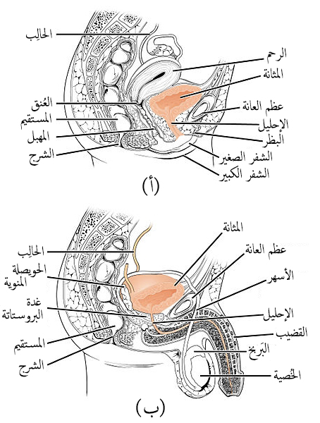
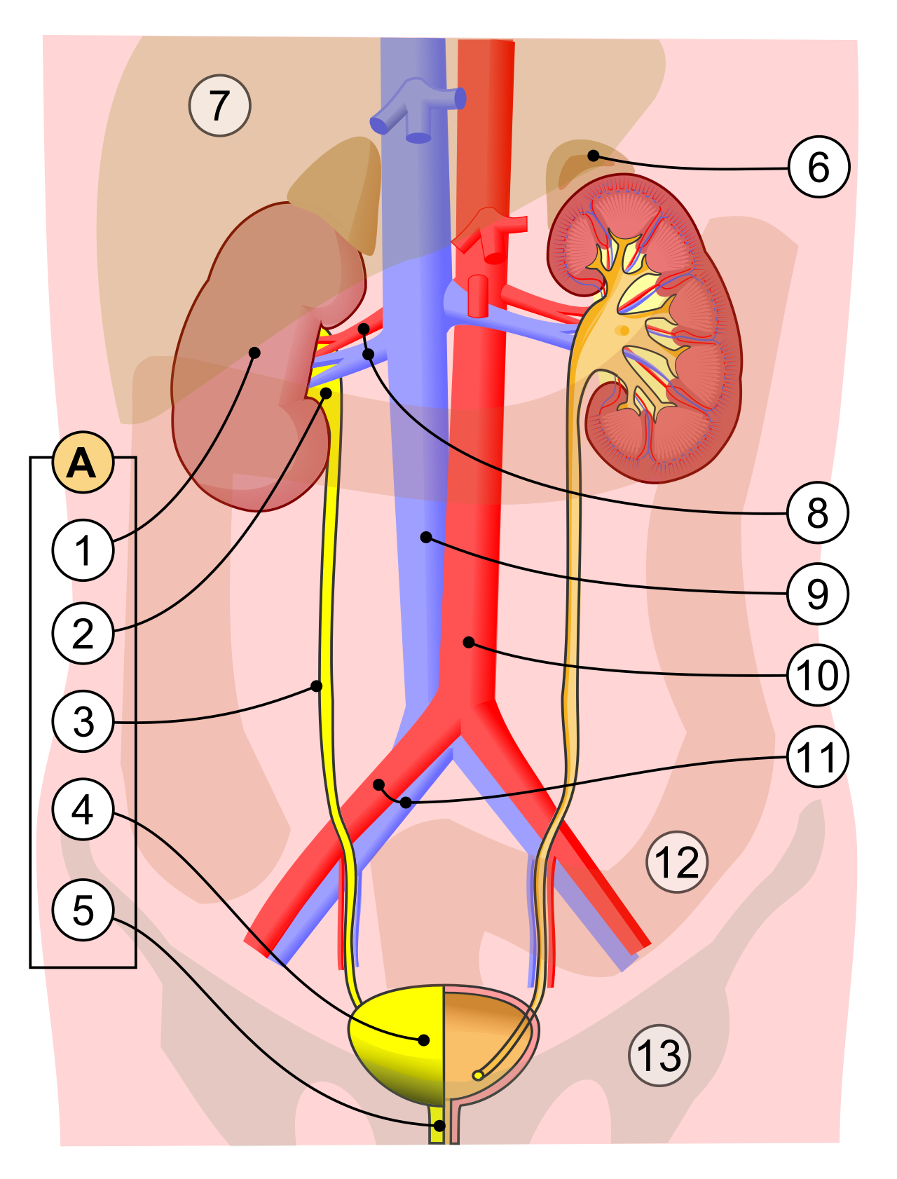
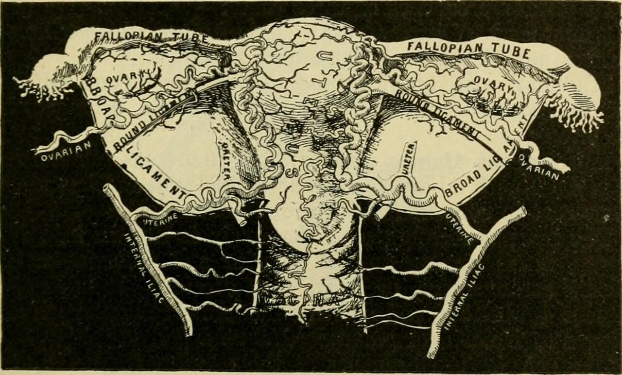
Description female pelvic cross section with numbered arrowsNo restrictions

By ruth lawson otago polytechnicCC BY 3.0

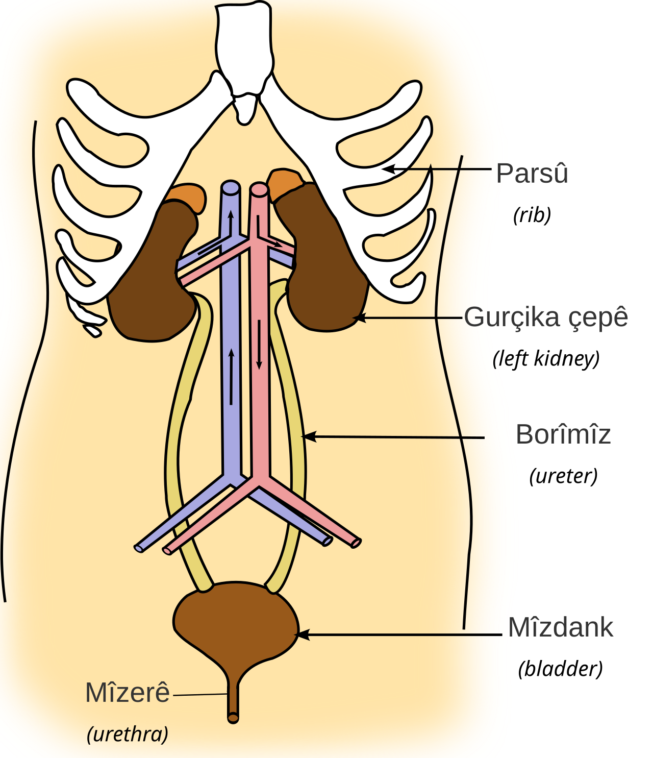
Diagram of urinary system unlabelled by ruth lawson otago poCC BY-SA 3.0

Title bladder and nearby organs female description the kidnePublic domain

Title bladder and nearby organs male description the kidneysPublic domain

Title digital rectal exam description digital rectal exam drPublic domain

Diagram of the formation of the human genito urinary apparatPublic domain
The urethra transports urine from the bladder to the outside

La uretra transporta la orina desde la vejiga hasta el exterCC BY 4.0

Diagrams to show the development of male and female generatiPublic domain

Diagrams to show the development of male and female generatiPublic domain

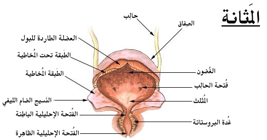
Kurdish koendama m z be a serek ya koendama derav tin ya mir

Plates 15 and 16 of roberton s the generative system 1824 dePublic domain

Anatomy of a common snailCC BY 3.0

BladderCC BY 4.0
Description female pelvic cross section with numbered arrowsNo restrictions
By ruth lawson otago polytechnicCC BY 3.0
Diagram of urinary system unlabelled by ruth lawson otago poCC BY-SA 3.0
Title bladder and nearby organs female description the kidnePublic domain
Title bladder and nearby organs male description the kidneysPublic domain
Title digital rectal exam description digital rectal exam drPublic domain
Diagram of the formation of the human genito urinary apparatPublic domain

The urethra transports urine from the bladder to the outside
La uretra transporta la orina desde la vejiga hasta el exterCC BY 4.0
Diagrams to show the development of male and female generatiPublic domain
Diagrams to show the development of male and female generatiPublic domain
Kurdish koendama m z be a serek ya koendama derav tin ya mir

Language neutral version of image illu urinary systemPublic domain

Title practical anatomy of the rabbit an elementary laboratoNo restrictions

Plates 15 and 16 of roberton s the generative system 1824 dePublic domain
Plates 15 and 16 of roberton s the generative system 1824 dePublic domain
Urinary system

Identifier anatomyphysiolog00jord find matches title anatomyNo restrictions

B yr k st v zil r lat glandulae suprarenales c t daxili sekrPublic domain

Identifier anatomydescripti1897gray find matches title anatoNo restrictions

Identifier anatomyphysiolog00walk find matches title anatomyNo restrictions
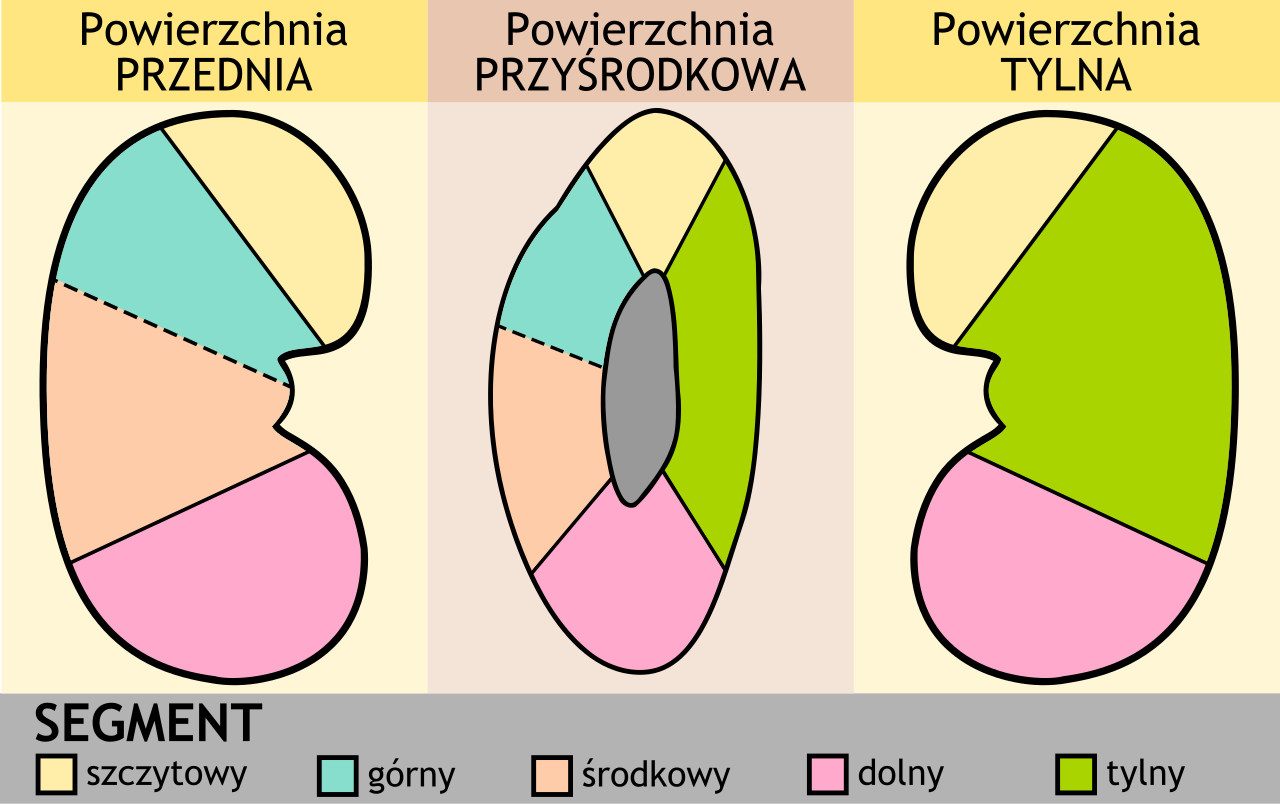
Arterial segments of right kidney
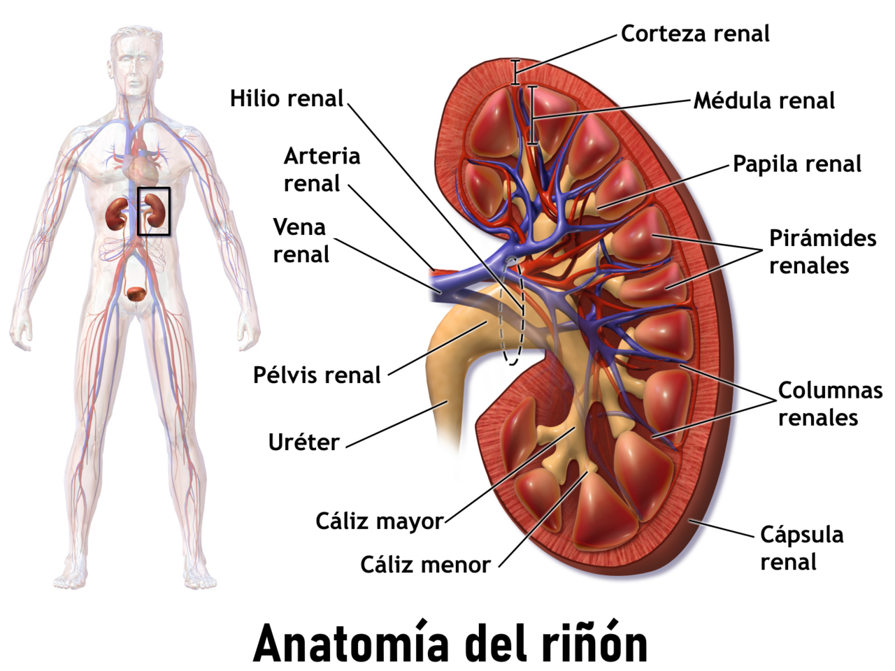
Anatom a del ri n vea una animaci n de este tema m dico

Title chordate anatomy identifier chordateanatomy00neal findNo restrictions

Diagram illustrating connexion between kidney and testis inPublic domain

A diagram of a hypothetical ancestral mollusc by nicholson aPublic domain

Schematic drawing of inner anatomy of a teleost fish 1 liverCC BY 2.5

Picture of kidneyPublic domain

Vertical section of kidney a version of file gray1127 png wiPublic domain

KidneyCC BY-SA 3.0

Language neutral version of image illu kidney2Public domain

Identifier b20416039 003 find matches title on the anatomy oNo restrictions

Identifier quainselementsofquai02 find matches title quain sNo restrictions

Identifier quainselementsofquai02 find matches title quain sNo restrictions

Identifier quainselementsof82quai02 find matches title quainNo restrictions

Diagram depicting two different renal hematomas and their reCC BY-SA 4.0

Illustration from anatomy amp physiology connexions web siteCC BY 3.0

Title anatomy in a nutshell a treatise on human anatomy in iNo restrictions

Title bensley s practical anatomy of the rabbit an elementarNo restrictions

Title comparative anatomy identifier comparativeanato00nealPublic domain

Title comparative anatomy of vertebrates identifier comparatNo restrictions

Skene s glands identifier diseasesofkidney01kell find matcheNo restrictions

Title elements of comparative anatomy identifier elementsofcNo restrictions
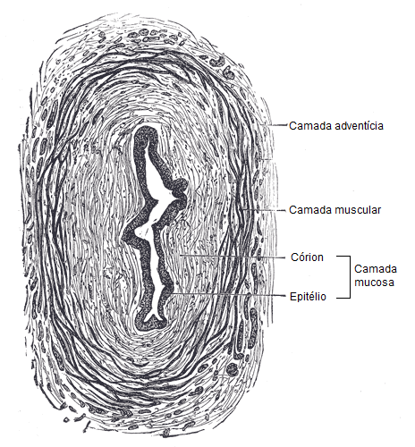
Sec o transversal do ureter mostrando suas estrutura histol
Identifier handbookofanatom00youn find matches title handbooInternet Archive Book Images

Illu ureters wallPublic domain

Title practical anatomy of the rabbit an elementary laboratoNo restrictions

Title practical anatomy of the rabbit an elementary laboratoNo restrictions

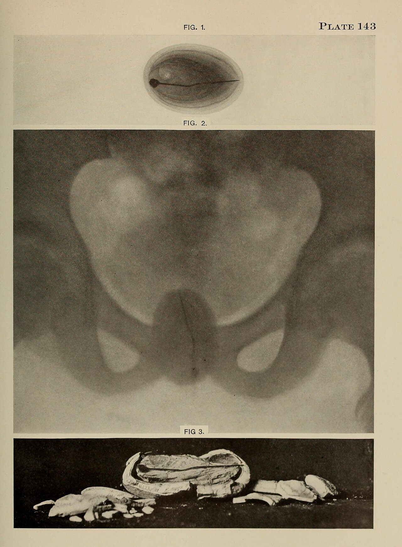
Identifier pyelographypyelo00braa find matches title pyelogrNo restrictions

Identifier pyelographypyelo00braa find matches title pyelogrNo restrictions

Identifier surgicalanatomyt02deav find matches title surgicaNo restrictions

Identifier americanjournalo13wist find matches title the ameNo restrictions

Title the anatomy of the domestic animals identifier anatomyNo restrictions

Title the anatomy of the domestic animals identifier anatomyNo restrictions

Title the anatomy of the domestic animals identifier anatomyNo restrictions

Title the anatomy of the domestic animals identifier anatomyNo restrictions

Title the anatomy of the domestic animals identifier anatomyNo restrictions

Title the anatomy of the domestic animals identifier cu31924No restrictions
BladderCC BY 4.0

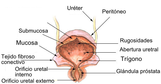
La vejiga urinariaCC BY 4.0

Illustration from anatomy amp physiology connexions web siteCC BY 3.0

Il lustraci anat mica de la bufeta de l orina les etiquetesCC BY 3.0
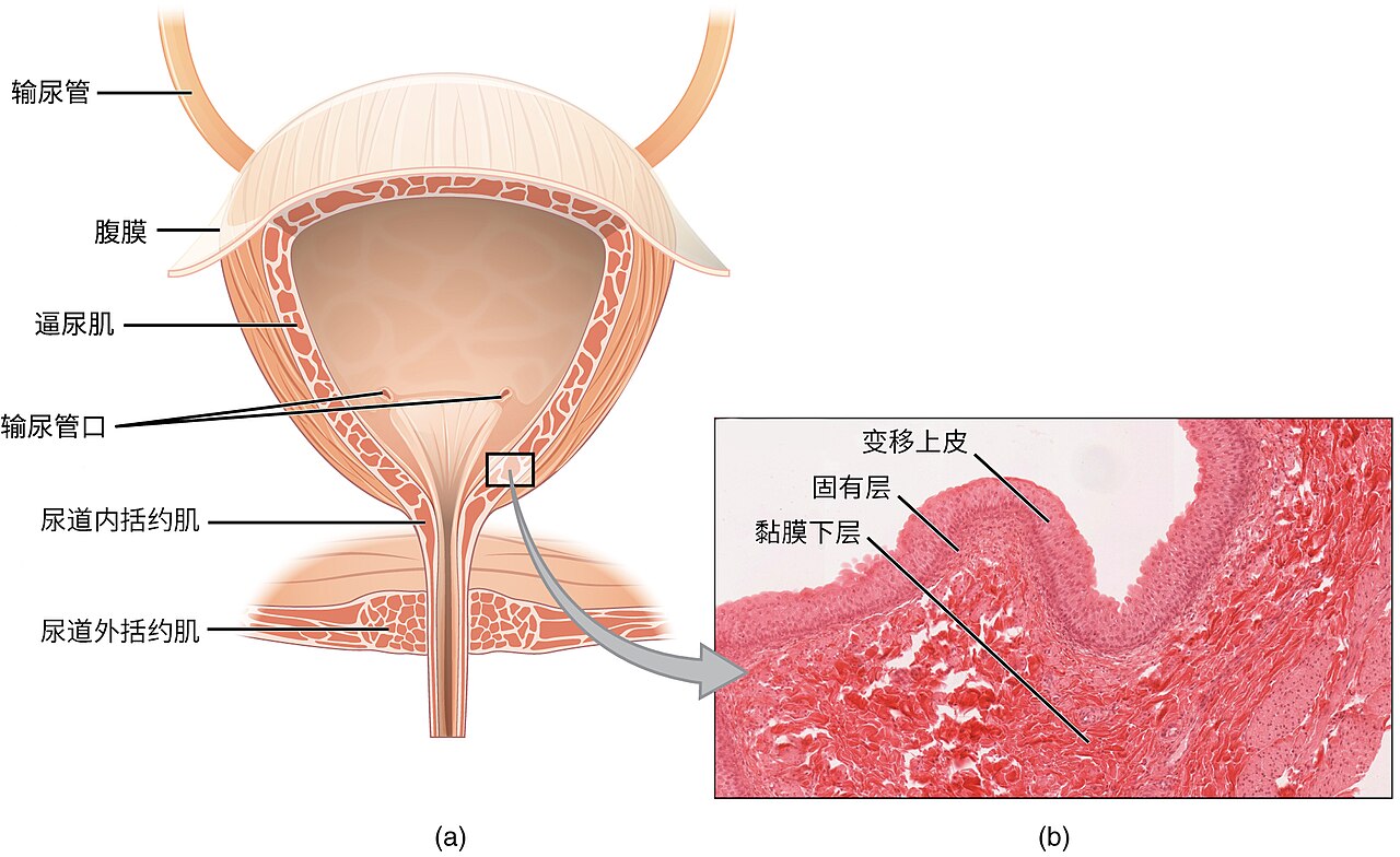
Illustration from anatomy amp physiology connexions web site

Identifier atlasofhumananat04tolduoft find matches title anNo restrictions

Woodcut illustration from an edition of 1537 16th year of jiCC BY 4.0

Unlabeled view of internal human urinary bladder anatomyCC BY-SA 4.0
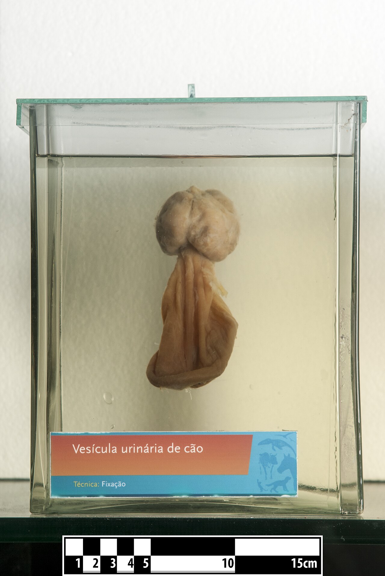
Dog urinary bladder canis lupus familiaris technique of form

View of the base of the bladder prostate seminal vesicles anPublic domain

Coronal section through the pelvis of a human male showing tPublic domain

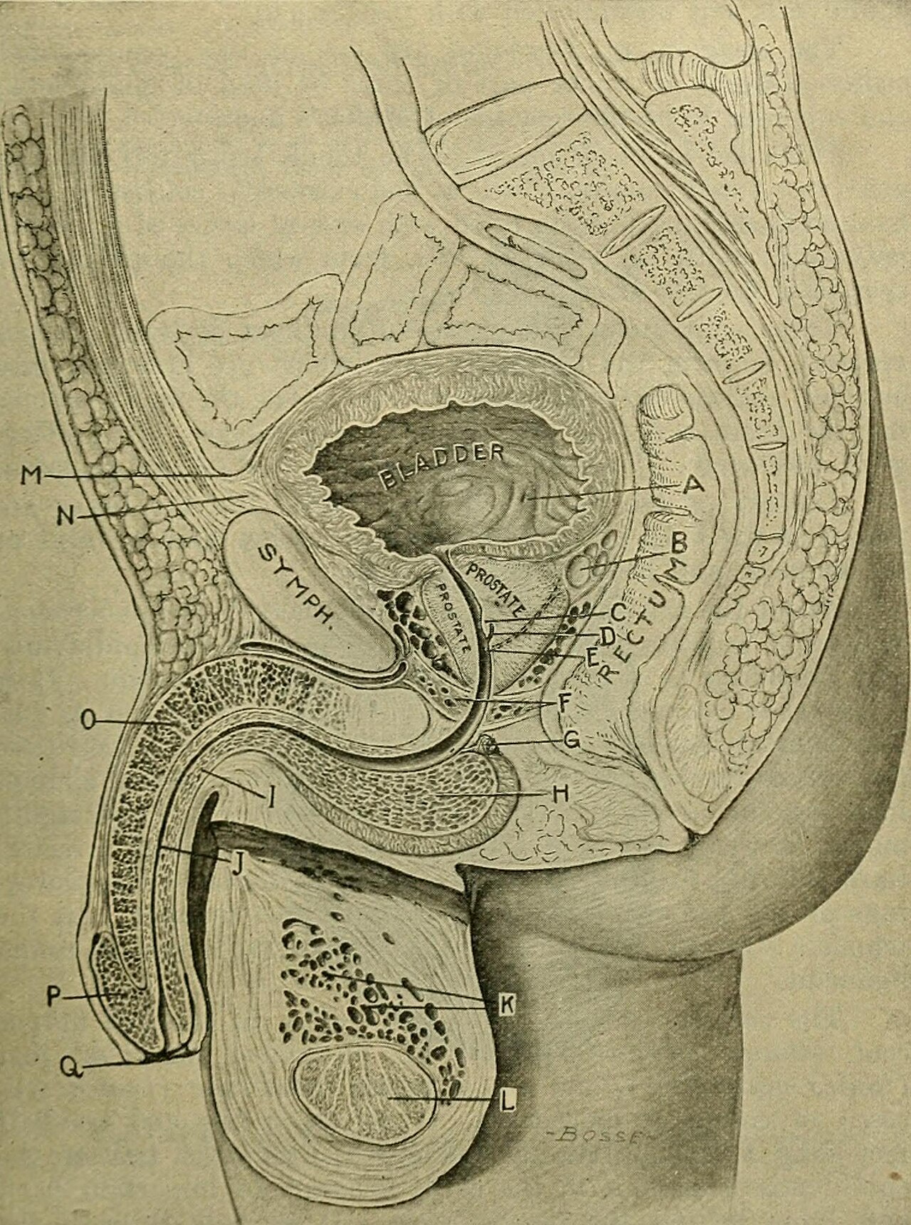
Sagittal median section of bladder prostate and rectum of aPublic domain
Anatomy of urinary bladder

Anatomy of urinary bladderPublic domain

Anatomy of urinary bladderPublic domain
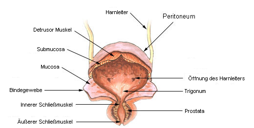
Aufbau der harnblase Translation by User:Lennert B
Anatomy of urinary bladder
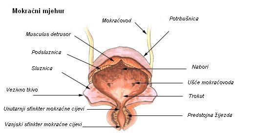
Anatomija mokra nog mjehura

Numbered version of illu bladderCC BY-SA 3.0
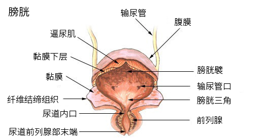
Anatomy of urinary bladder

Position of the urinary bladder in man and womanCC BY-SA 4.0
Neck of the bladder identifier anatomyphysiolog03belluoft fiInternet Archive Book Images

Identifier surgicaldiseases00keye find matches title the surNo restrictions

Fig 1018 the urinary bladder prostate and seminal vesicle viPublic domain

Version 8 25 from the textbook openstax anatomy and physioloCC BY 4.0

Muscles of the perineum identifier treatiseonpracti00boen fiNo restrictions

Identifier treatiseonpracti00boen find matches title a treatNo restrictions

Identifier treatiseonpracti00boen find matches title a treatNo restrictions

Identifier atlasofhumananat04tolduoft find matches title anNo restrictions

Fig 6 analysis of the urethra in c57bl 6j male mice identifiCC BY 4.0
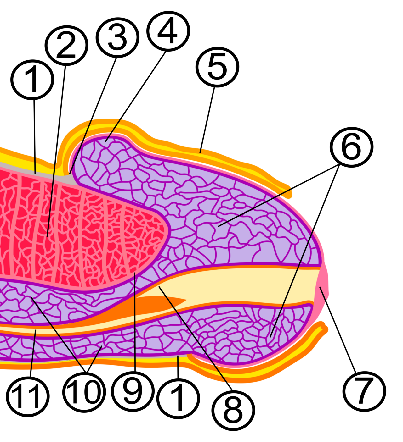
Internal anatomy of human glans penis 1 fascia penis 2 corpu drawing, adaptation and modification by Avereanu

Identifier clinicallectures00harr find matches title clinicaNo restrictions

Instrument introduced into the urethra takes incorrect pathNo restrictions

External vagina urethra and anus rare books keywords williamCC BY 4.0

The urethra transports urine from the bladder to the outsideCC BY 3.0
Identifier handbookofanatom00youn find matches title handbooInternet Archive Book Images
Identifier handbookofanatom00youn find matches title handbooInternet Archive Book Images
Stone in urethra encapsulating a pin girl 13 years old upperRotch, Thomas Morgan

Male urethral meatus at the tip of the glans penisCC0

Identifier manualofpatholog00jone find matches title manualNo restrictions

Zeichnung der m nnlichen anatomisch aufgeschnittenen urethraPublic domain

Identifier surgicalanatomyt02deav find matches title surgicaNo restrictions
Title the anatomy of the horse a dissection guide identifierInternet Archive Book Images

Nephron filtration der freien leichten immunglobulinkettenCC BY-SA 3.0

Diagram shows the sequential development and degeneration ofCC BY-SA 3.0

Diagram showing how the kidneys workCC BY-SA 4.0

Diagram showing human nephron with pct dct and henle s loopCC BY-SA 4.0

ImageCC BY-SA 3.0
Schematics of kidney and nephron 1 renal cortex 2 medulla 3

This images shows the cells that make up a renal corpuscle oCC BY 3.0

Nefr diagrama de la formaci de l orina el nombre de dins delCC BY-SA 3.0

ImageCC BY-SA 3.0
Nephron diagram of the urine formation the number inside tub
Nephron diagram of the urine formation the number inside tub

Unlabelled diagram of a mammalian nephronCC BY-SA 4.0

Diagram of filtration secretion and reabsorption in nephronCC BY-SA 4.0

Basic anatomy of nephronCC BY 3.0
Physiology of nephrons

Physiology of nephronCC BY 3.0

Fisiolog a de la nefronaCC BY-SA 3.0

Nefronaren fisiologiaCC BY-SA 3.0

Kurdish m z ji p khatey n nav plazmaya xw n di nefron n gurCC BY 3.0
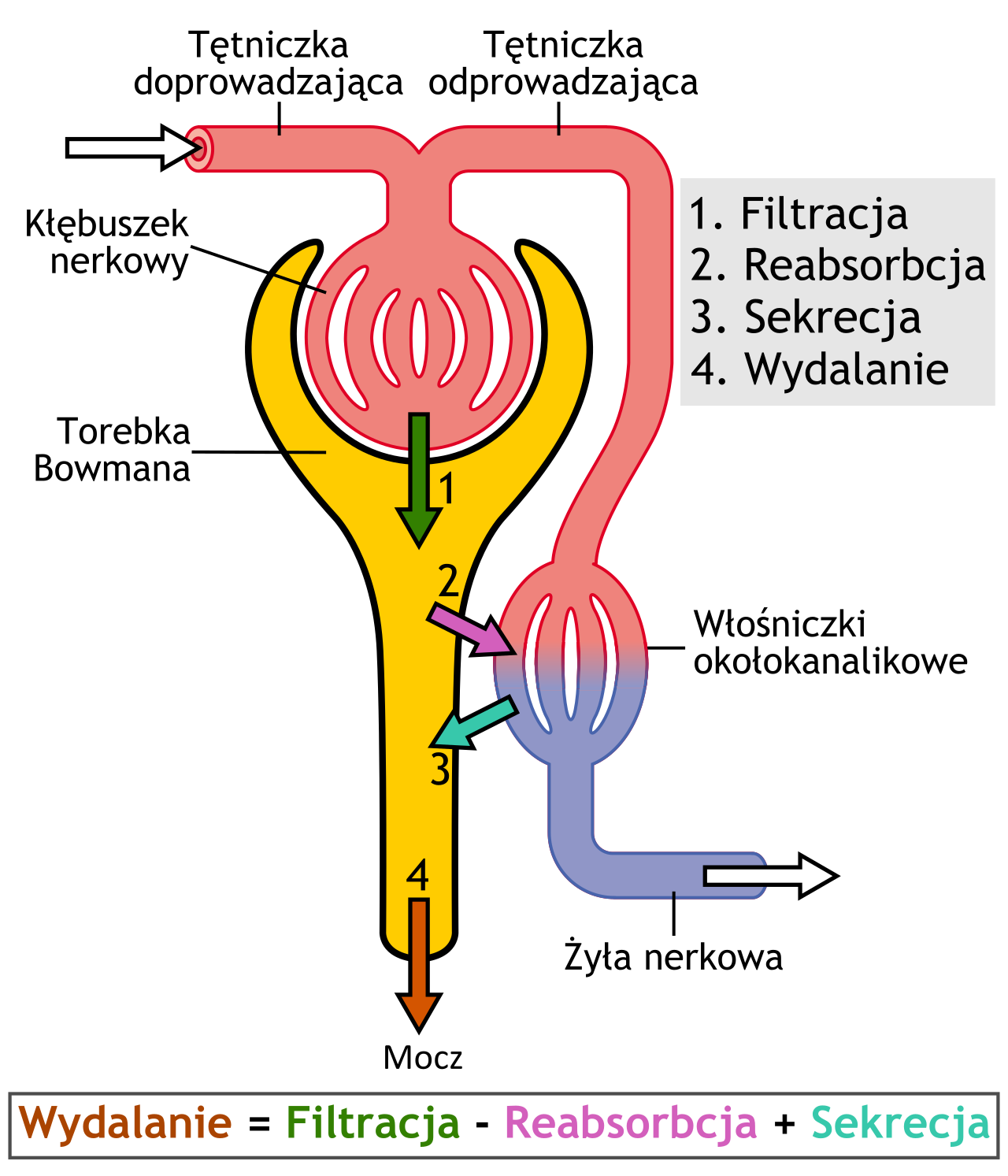
Physiology of nephron
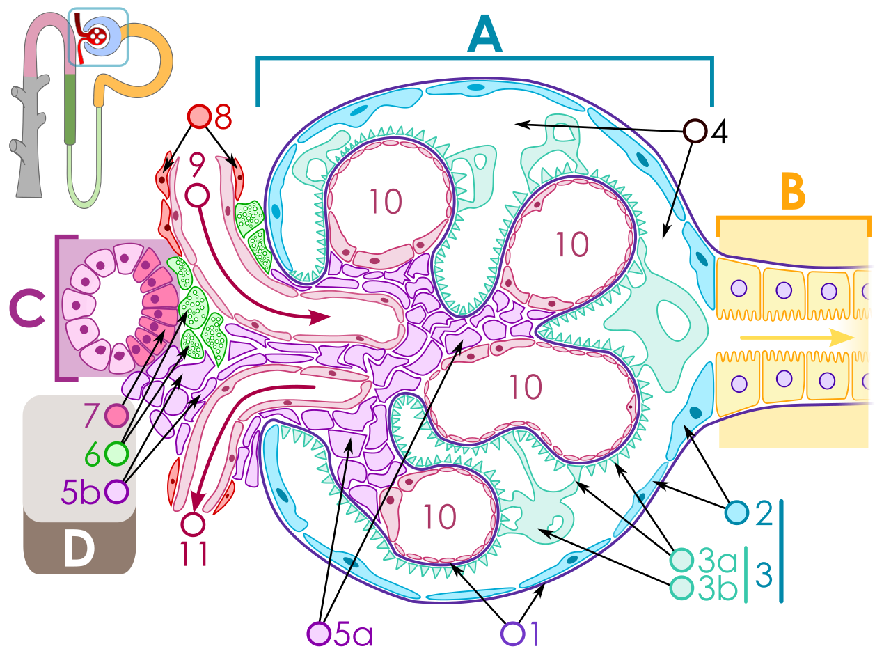
Diagram of renal corpuscle structure a renal corpuscle b pro

Identifier 2578007rx2 nlm nih gov title a system of anatomyNo restrictions

Title anatomy descriptive and applied identifier anatomydescNo restrictions
Identifier anatomydescripti1897gray find matches title anato

Identifier anatomyphysiolo00hewe find matches title anatomyNo restrictions

Title anatomy of the cat identifier anatomyofcatrje00reig fiNo restrictions
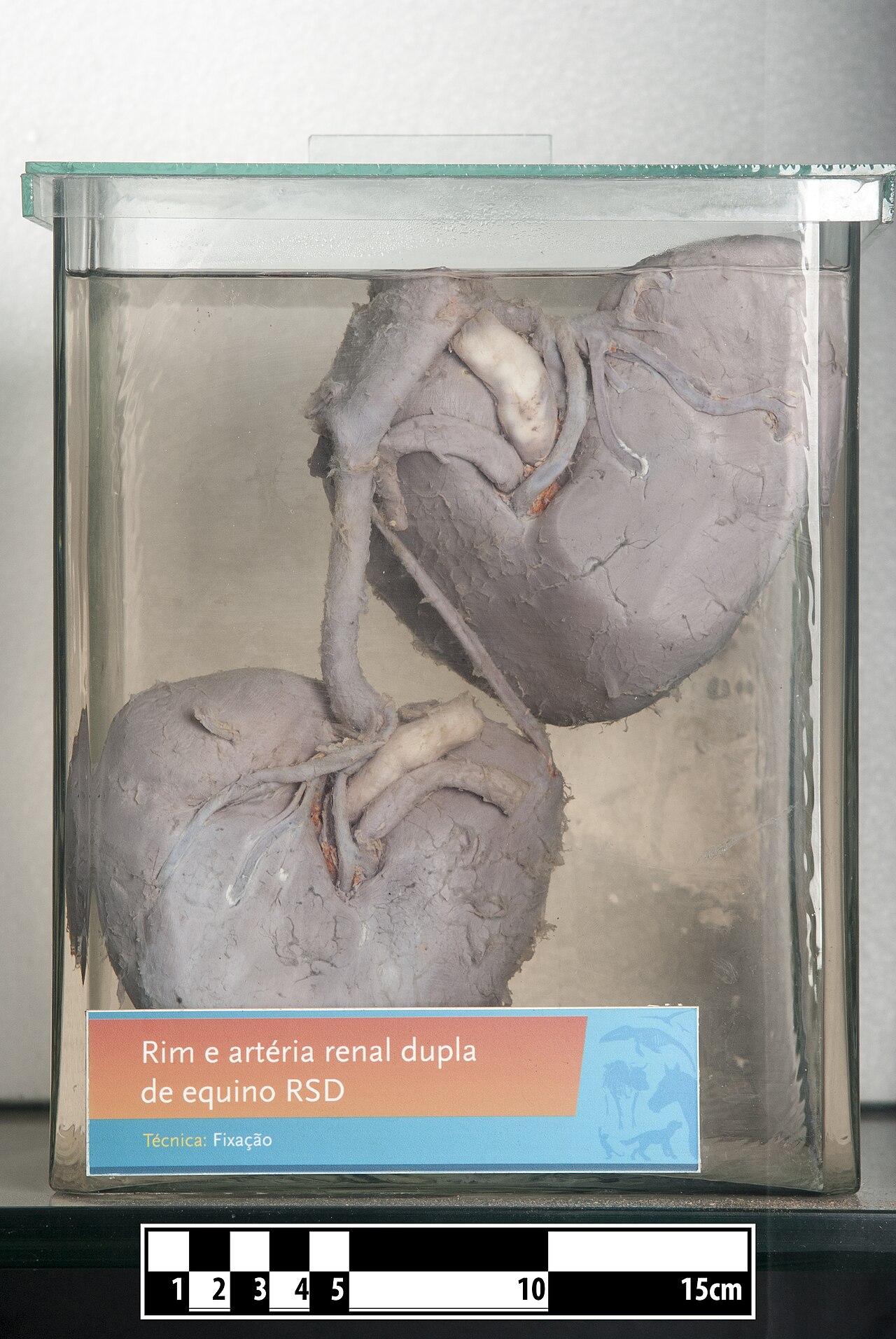
Horse kidneys with renal artery technique of formalin fixati

Identifier manualofpatholog00jone find matches title manualNo restrictions

Identifier quainselementsof82quai02 find matches title quainNo restrictions

Identifier quainselementsof82quai02 find matches title quainNo restrictions

Identifier quainselementsofquai02 find matches title quain sNo restrictions

Renal arteries rare books keywords anatomy albrecht von hallCC BY 4.0

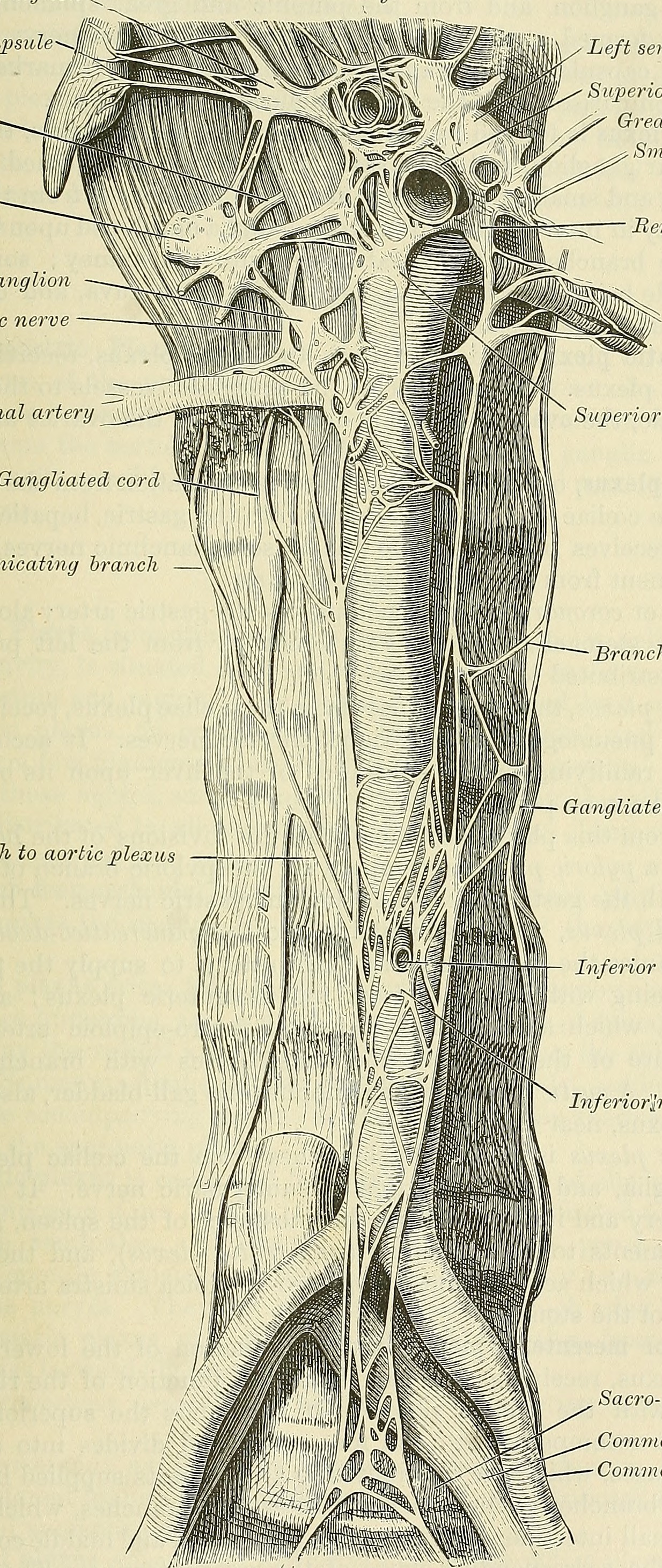
Renal system arteries etc wellcome images keywords anatomyCC BY 4.0

Title the anatomy of the domestic animals identifier anatomyNo restrictions

Title the anatomy of the domestic animals identifier cu31924No restrictions

Title the anatomy of the domestic animals identifier cu31924No restrictions

Title the comparative anatomy of the domesticated animals idNo restrictions

Title the comparative anatomy of the domesticated animals idNo restrictions

Title the comparative anatomy of the domesticated animals idNo restrictions

Title the comparative anatomy of the domesticated animals idNo restrictions

Title the comparative anatomy of the domesticated animals idNo restrictions

Title the cyclop dia of anatomy and physiology identifier cyNo restrictions

Title the cyclop dia of anatomy and physiology identifier cyNo restrictions

Identifier operativegynecol001kell find matches title operatNo restrictions

Identifier systemri01hirs find matches title a system of obsNo restrictions

Nine diagrams illustrating cross sections through the male rCC BY 4.0
Anatomie - appareil genital masculin
115 images
Title a laboratory manual for comparative vertebrate anatomyNo restrictions

Anatom a del aparato genital masculinoCC BY 4.0

Title chordate anatomy identifier chordateanatomy00neal findPublic domain

Diagram to illustrate the structure of the testis and epididPublic domain
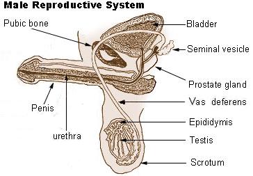
Diagram of a cross section of the human male reproductive sy original uploader was NightFlyer at en.wikipedia.

Title comparative anatomy of vertebrates identifier comparatNo restrictions

Internal male reproductive system in lethocerus patruelis tCC BY 3.0

Male genital system of a domestic rabbitCC BY-SA 3.0

M nnliche genitalorganeCC BY-SA 4.0

Title the chordates identifier chordates00rand find matchesNo restrictions

Identifier b20388615 find matches title a manual of dissectiNo restrictions

Title anatomy descriptive and applied identifier anatomydescNo restrictions

Title anatomy descriptive and applied identifier anatomydescNo restrictions

Ki i xarici cinsiyy t zv penis sidik kanal prostat v zi meriCC BY-SA 3.0

Two panel drawing showing normal male reproductive and urinaCC BY-SA 4.0
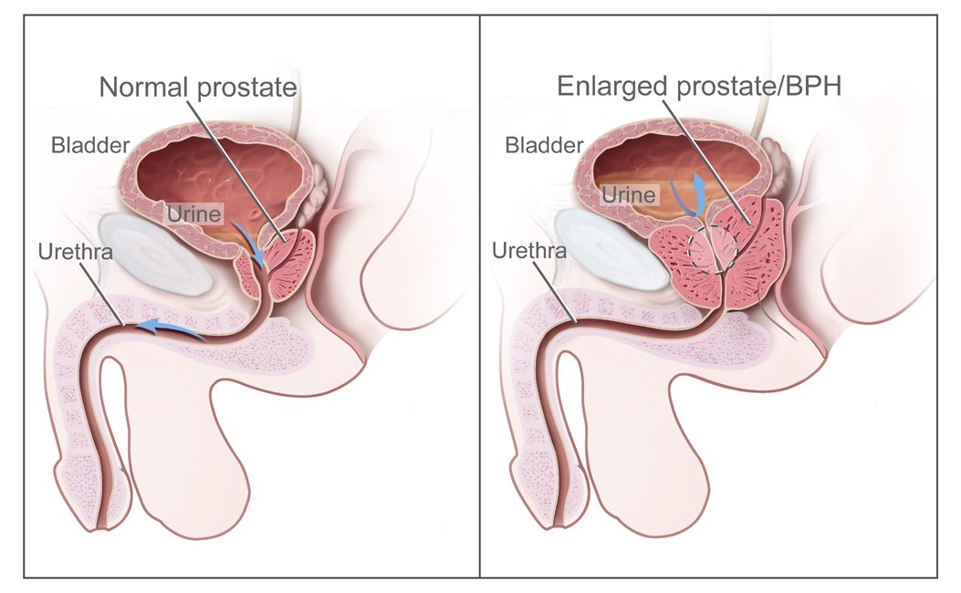
Title benign prostatic hyperplasia description two panel dra

Title comparative anatomy identifier comparativeanato00nealPublic domain

Title comparative anatomy of vertebrates identifier comparatNo restrictions

Title cunningham s text book of anatomy identifier cunninghaNo restrictions

Title cunningham s text book of anatomy identifier cunninghaNo restrictions
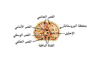
Illu prostate lobes ar
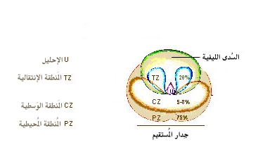
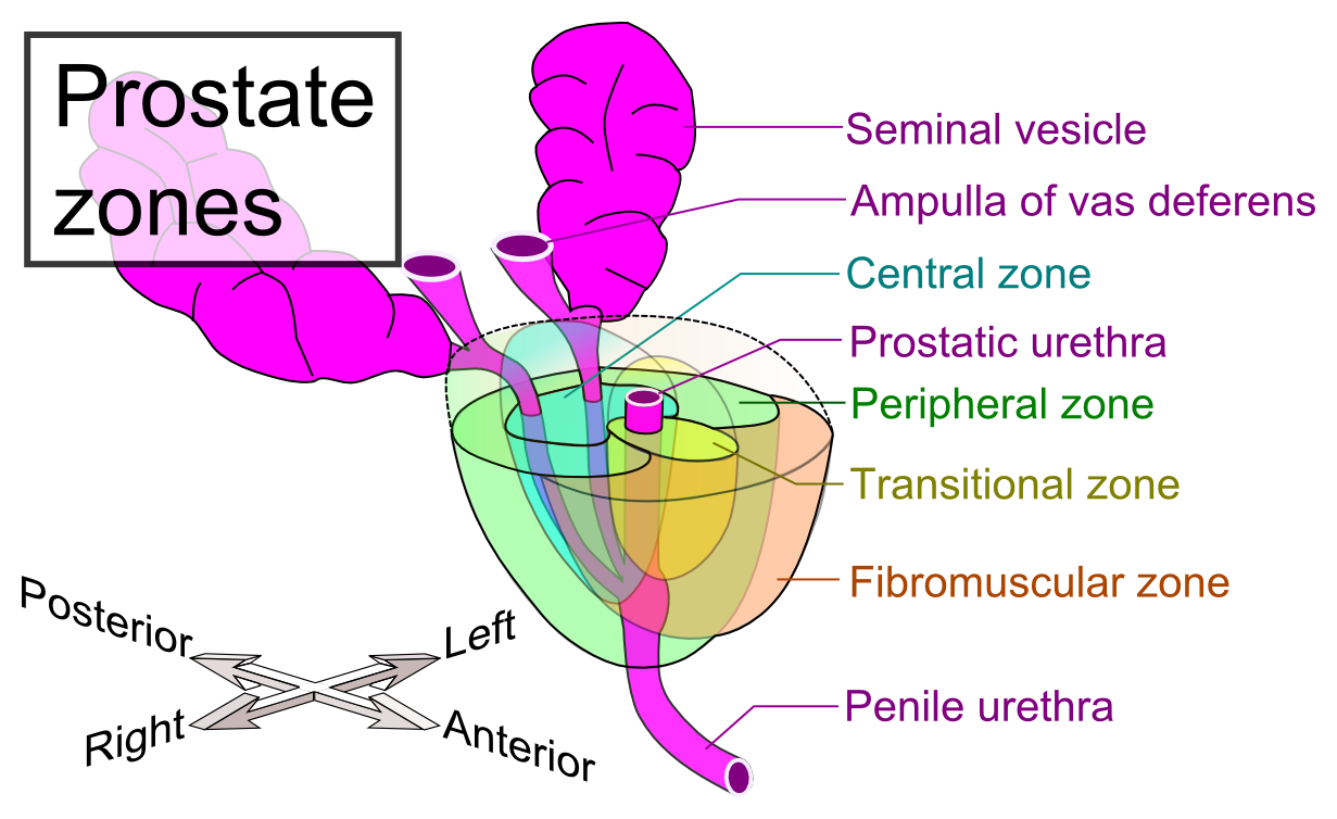
Illu prostate zones ar

Title an introduction to the study of the comparative anatomNo restrictions

The female human genitalia soruce for the lettering of the pCC0

Identifier americanjournalo13wist find matches title the ameNo restrictions

Identifier americanjournalo13wist find matches title the ameNo restrictions

Title the chordates identifier chordates00rand find matchesNo restrictions

Title the comparative anatomy of the domesticated animals idNo restrictions

Toucher rectal 1 vessie 2 rectum 3 prostateCC BY-SA 3.0

Identifier atlasofhumananat04tolduoft find matches title anNo restrictions

Title an atlas of human anatomy for students and physiciansNo restrictions

Title comparative anatomy identifier comparativeanato00nealPublic domain

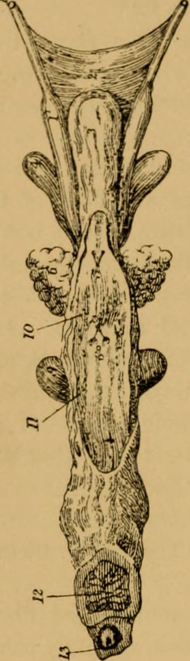
Fig 1015 diagram to illustrate the descent of the testis andPublic domain

Schematic drawing of testicle and epidydimis 1 tunica albugiCC BY 2.5

Ilustraci n de las partes de un test culoCC BY 4.0

Illu testis cross sectionPublic domain

Illu testis surfacePublic domain

A diagram of the major components of an adult human testicleCC BY-SA 3.0

Identifier anatomistsvademe1851wils find matches title the aNo restrictions

Identifier atlasofhumananat04tolduoft find matches title anNo restrictions

Anatomical diagram of a circumcised penis on a middle aged cCC BY-SA 3.0

Anatomical diagram of a circumcised penis on a middle aged cCC BY-SA 3.0

This is an annotated image of the glans penis of a human aloCC0
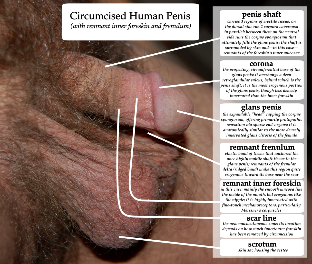
A diagram of a circumcised human penis with remnant inner fo

Diagram of penis with penile raphe clearly definedCC BY-SA 4.0

Diagram showing the anatomy of the penisCC BY-SA 4.0

Vaginalverkehr bezeichnung der u eren genitalorganeCC BY-SA 4.0

Diagram on the anatomy of a male mammal human penis not inclPublic domain
Illu penis ar

Anatomy of human penis parts numberedCC BY-SA 3.0

Distribution of penis circumference in inchesCC BY-SA 3.0

Cumulative distribution of penis circumference in inchesCC BY-SA 3.0

Distribution of penis length in inchesCC BY-SA 3.0

Results from lifestyles condom manufacturer s study of penisCC BY-SA 3.0

Identifier treatiseonpracti00boen find matches title a treatNo restrictions

Title anatomy descriptive and applied identifier anatomydescNo restrictions

Title anatomy in a nutshell a treatise on human anatomy in iNo restrictions

Title anatomy in a nutshell a treatise on human anatomy in iNo restrictions

Title chordate anatomy identifier chordateanatomy00neal findNo restrictions

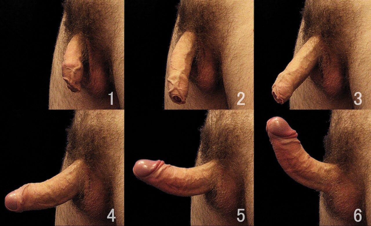
Photograph of a human male penis in an erect state the scrotCC BY-SA 3.0

The photograph illustrates the male external reproductive orCC BY 4.0

Illustration from anatomy amp physiology connexions web siteCC BY 3.0

Scrotum after it has been prepared for selling as souvenirCC BY-SA 4.0

Lateral and dorsal view human penis and scrotumCC0

Lateral view of human penis and scrotumCC0

Male genital anatomy penis erection states a relaxation phasPublic domain

Male genital anatomy penis flaccid originalCC BY-SA 3.0

Male genital anatomy scrotum and perineum close upCC BY-SA 3.0

Male genital anatomy scrotum and perineum close up 01 scrotuPublic domain

This photo shows the difference between a relaxed and tenseCC BY-SA 4.0

Scrotum du boucCC BY 4.0

Shaved human penis scrotum perineum and anus with labelsCC0

Identifier surgicalanatomyt02deav find matches title surgicaNo restrictions

Identifier surgicalanatomyt02deav find matches title surgicaNo restrictions

Identifier surgicalanatomyt02deav find matches title surgicaNo restrictions

Title anatomy in a nutshell a treatise on human anatomy in iNo restrictions

Title anatomy in a nutshell a treatise on human anatomy in iNo restrictions

Title anatomy in a nutshell a treatise on human anatomy in iNo restrictions

Identifier anatomyofarterie00powe find matches title anatomyNo restrictions

Some of male reproductive organs testicles testis spermaticCC BY-SA 3.0

Anatomical preparationCC BY-SA 3.0

Anatomical preparationCC BY-SA 3.0

Anatomical preparationCC BY-SA 3.0

Anatomical preparationCC BY-SA 3.0

Anatomical preparationCC BY-SA 3.0

Identifier surgicalanatomyt02deav find matches title surgicaNo restrictions

Identifier surgicalanatomyt02deav find matches title surgicaNo restrictions

Title the anatomy of the domestic animals identifier anatomyNo restrictions

Title the anatomy of the domestic animals identifier anatomyNo restrictions

Title the anatomy of the domestic animals identifier anatomyNo restrictions

Title the anatomy of the domestic animals identifier cu31924No restrictions

Title the anatomy of the domestic animals identifier cu31924No restrictions

By ruth lawson otago polytechnicCC BY 3.0

An illustration of an adult human testicle with the epididymCC BY-SA 3.0

Kurdish p khateya gun alozeyCC BY-SA 3.0

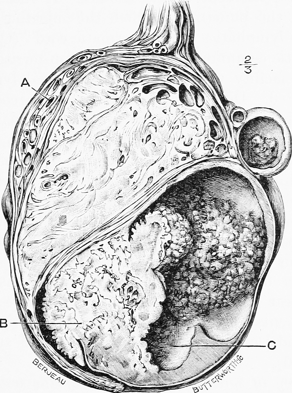
The right testis exposed by laying open the tunica vaginalisPublic domain

Fig 1007 left testis and epididymis viewed from behind showiPublic domain

Identifier b20416039 003 find matches title on the anatomy oNo restrictions

Identifier surgicalanatomyt02deav find matches title surgicaNo restrictions

Adapted from https commons wikimedia org wiki file testis pnCC BY-SA 4.0

Title the cyclop dia of anatomy and physiology identifier cyNo restrictions

Title an american text book of physiology identifier americaNo restrictions

Title animal biology identifier animalbiology00wood find matNo restrictions

Title biology of the vertebrates a comparative study of manNo restrictions
Fig 3 schematic diagram of copulatory apparatuses in chimaer Hagiya (2023)

Dendrocoelopsis piriformis diagram of the copulatory organsPublic domain

Polycelis borealis diagram of the copulatory organs in longiPublic domain

Diagram depicting the location of various organs in sexual sCC BY 2.5

Identifier journalofexperim31harr find matches title the jouNo restrictions

Title biology identifier biologycalk00calk find matches yearNo restrictions

Dendrocoelopsis alaskensis diagram of the copulatory organsPublic domain

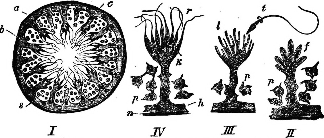
Fig 6 diagram of the formation of the genito urinary apparatPublic domain

Phagocata nivea diagram of the copulatory organs in longitudPublic domain

Title the veterinary bacteriological laboratories issued inNo restrictions
Figures 4 8 triannulata magna fig 4 adult in latero ventral Gelder, S.R. (2020). North American Branchiobdellida (Annelida: Clitellata) or Crayfish Worms in France: the most diverse distribution of these exotic ectosymbionts in Europe. Zoosymposia, 17: 121–140.
Lithiase urinaire
79 images
A man with kidney stones suffering from the typical symptomCC BY-SA 4.0
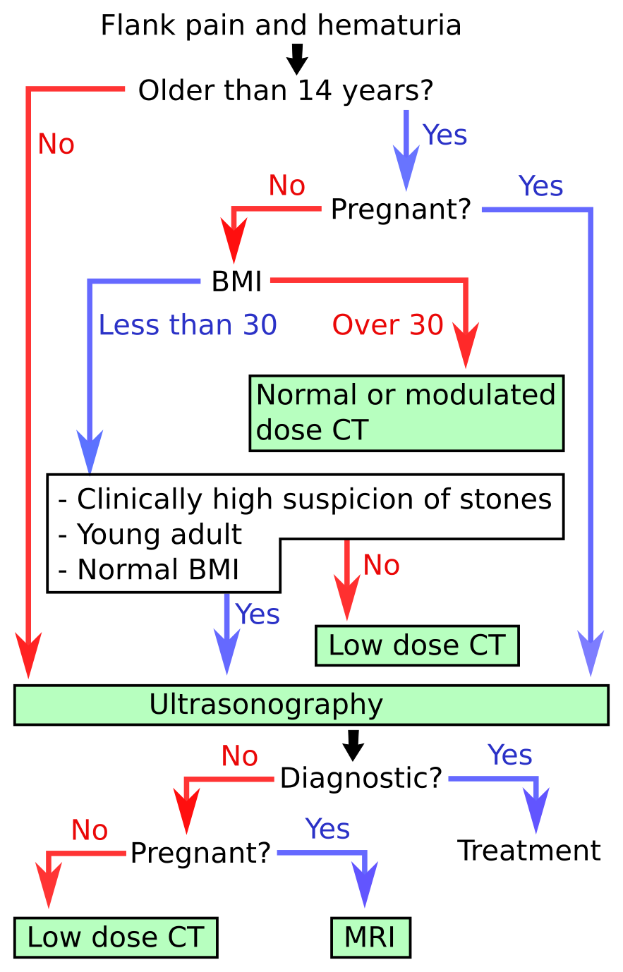
An algorithm for the diagnosis of kidney stone disease

Ct kub scan used for detailed imaging of kidneys ureters andCC BY 4.0

Kidney stone that i passed naturally albeit painfullyCC BY 2.5

Title ultrasonic instrument and kidney stone image id 4172 pPublic domain

A lenticular kidney stone excreted by the urine this is an eCC BY-SA 4.0

A kidney stoneCC0

A kidney stone yet unidentified type author supposed calciumCC BY-SA 3.0

Kidney stone 4mmCC0

Kidney stone 4mmCC0

Kidney stone 4mmCC0

Kidney stone 4mmCC0

Kidney stone 4mmCC0

Kidney stone with a maximum dimension of 5mmPublic domain

These are some of the larger passed fragments of a 1 cm kidnPublic domain

A dark kidney stone is excreted in a woman s urine in her 60CC BY-SA 4.0

Kidney stones successfully removed from an elderly dog codyPublic domain

Kidney stones one entire and other in small pieces after extCC BY-SA 4.0

A lenticular kidney stone excreted by the urineCC BY-SA 4.0

Kidney stonePublic domain
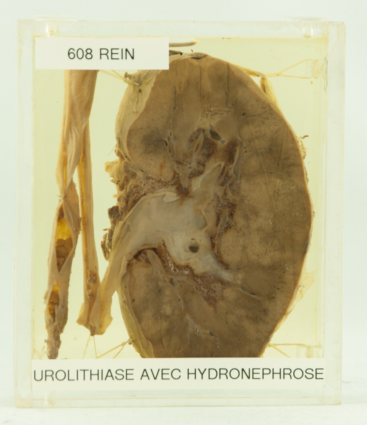
Diagnostic urolithiase avec hydron phrose histoire femme de
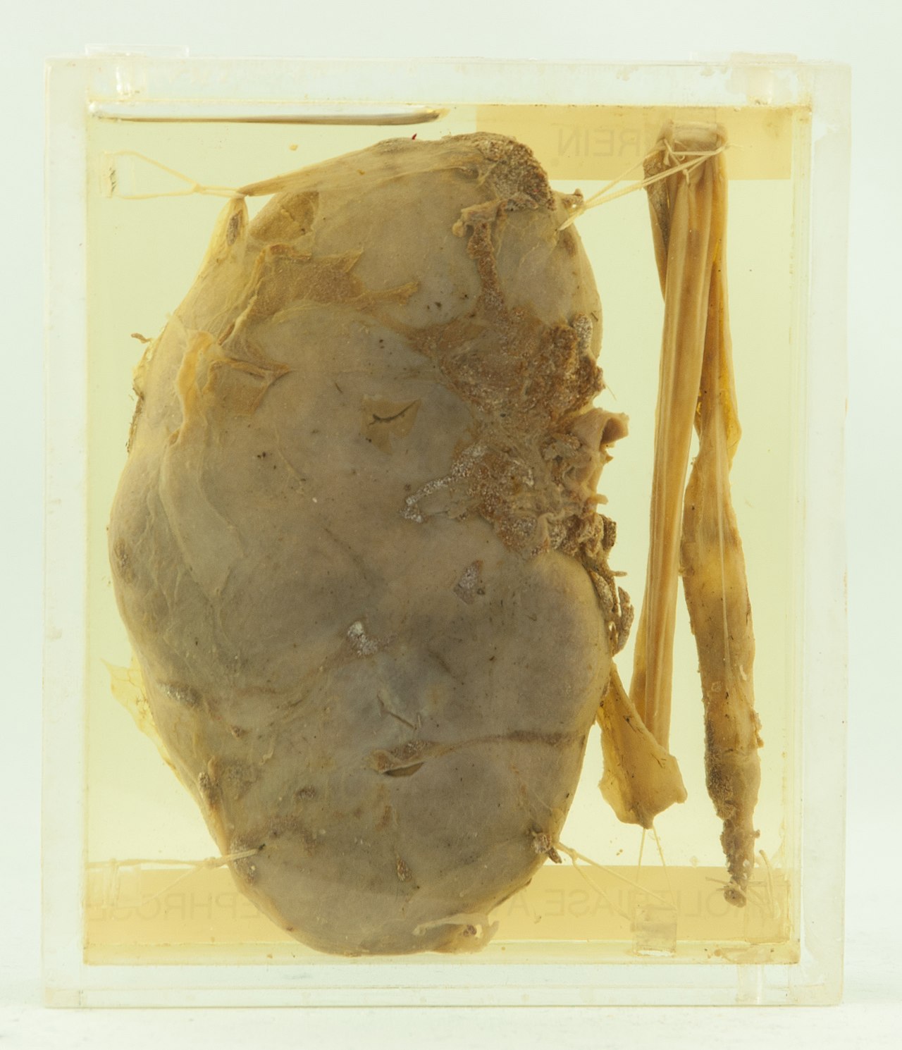
Diagnostic urolithiase avec hydron phrose histoire femme de

A renal stone from renal pelvis stag horn like stoneCC BY-SA 3.0
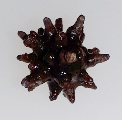
A 1 cm big spiculated kidney stone resembling a morning star

Small crystals formed in kidney the most common renal calculCC BY-SA 3.0

Calcium deposits in renal parenchyma note partial staghorn cCC0

Bilateral kidney stones on abdominal x ray not to be confuseCC BY 2.0

Passing a kidney stone can sometimes happen free of symptomsCC BY-SA 4.0

Ultrasound of left kidney lower pole with stone and posterioCC BY-SA 4.0

Identifier annualreportofst19142mary find matches title annuNo restrictions

A giant ureter stoneCC BY 4.0

Fig 1 cut to illustrate the common location of ureteral calcNo restrictions

Classificazione dei calcoli delle vie urinarie per posizioneCC BY-SA 4.0

Cut to illustrate the common location of ureteral calculus ePublic domain

Identifier diseasesofkidney01kell find matches title diseaseNo restrictions

Identifier diseasesofkidney01kell find matches title diseaseNo restrictions

Identifier diseasesofkidney01kell find matches title diseaseNo restrictions

Identifier diseasesofkidney01kell find matches title diseaseNo restrictions

Identifier diseasesofkidney01kell find matches title diseaseNo restrictions

Identifier diseasesofkidney01kell find matches title diseaseNo restrictions

Identifier interstatemedica2419unse find matches title interNo restrictions

Created by morning2k a kub film shows an radiopaque lesion oCC BY 2.5

Identifier onoriginprogress00morr find matches title on theNo restrictions

Identifier onoriginprogress00morr find matches title on theNo restrictions

Identifier onoriginprogress00morr find matches title on theNo restrictions

Identifier operativegynecol001kell find matches title operatNo restrictions

Hydronephrosis due to a kidney stone at the ureteral vesiculCC BY-SA 3.0

A stone at the ureteral vesicular junctionCC BY-SA 3.0

3d medical animation still showing ureteral stonesCC BY-SA 4.0

Identifier transactionsofso2319sout find matches title transNo restrictions

Three dimensional reconstructed ct scan image of a ureteralCC BY-SA 3.0

Identifier virginiamedicals2119unse find matches title virgiNo restrictions

Kidney stonesCC BY-SA 4.0

Kidney stonesCC BY-SA 4.0

Renal calculi identifier textbookofpracti1885loom find matchNo restrictions

Alexander marcet 1770 1822 was a genevan physician who speciPublic domain

Alexander marcet 1770 1822 was a genevan physician who speciPublic domain

Alexander marcet 1770 1822 was a genevan physician who speciPublic domain

Alexander marcet 1770 1822 was a genevan physician who speciPublic domain

Alexander marcet 1770 1822 was a genevan physician who speciPublic domain

Alexander marcet 1770 1822 was a genevan physician who speciPublic domain

Alexander marcet 1770 1822 was a genevan physician who speciPublic domain

Alexander marcet 1770 1822 was a genevan physician who speciPublic domain

Alexander marcet 1770 1822 was a genevan physician who speciPublic domain

Alexander marcet 1770 1822 was a genevan physician who speciPublic domain

Alexander marcet 1770 1822 was a genevan physician who speciPublic domain

Alexander marcet 1770 1822 was a genevan physician who speciPublic domain

Alexander marcet 1770 1822 was a genevan physician who speciPublic domain

Alexander marcet 1770 1822 was a genevan physician who speciPublic domain

Alexander marcet 1770 1822 was a genevan physician who speciPublic domain

Alexander marcet 1770 1822 was a genevan physician who speciPublic domain

Calcium carbonate crystals are a type of urinary sediment anCC BY-SA 4.0

Le griffon de la source cachat avec la nymphe la source la sCC BY 2.0

Kidney and bladder stones from authors collection and that oPublic domain

Identifier practicalelectro00mart find matches title practicNo restrictions

Identifier rntgenrayselectr00kass find matches title r ntgenNo restrictions

Identifier roentgenraysinme1901will find matches title the rNo restrictions

Ct of abdomen without contrast showing right proximal ureterCC BY-SA 3.0

A kidney stone with associated hydronephrosis mid ureterCC BY-SA 3.0
Cancerologie - rein
99 images
From a slide study set pathological and histological imagesCC BY 2.0
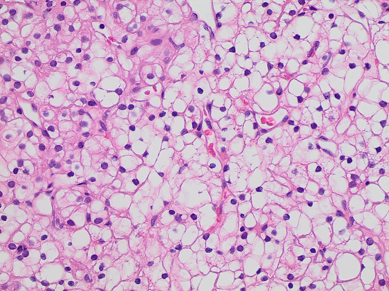
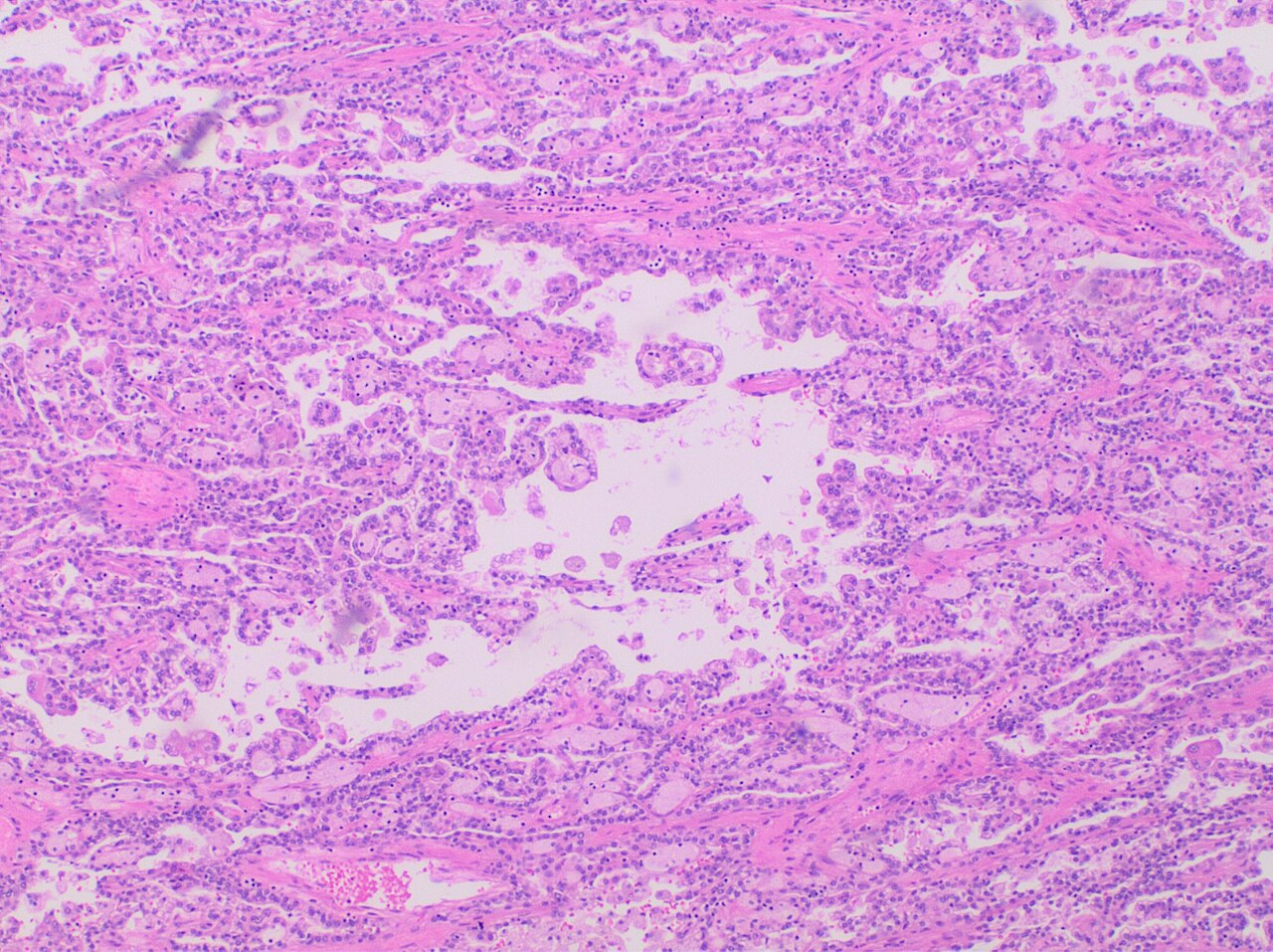
Grade 2 clear cell renal cell carcinoma showing clearly visi
Grade 2 clear cell renal cell carcinoma showing clearly visi

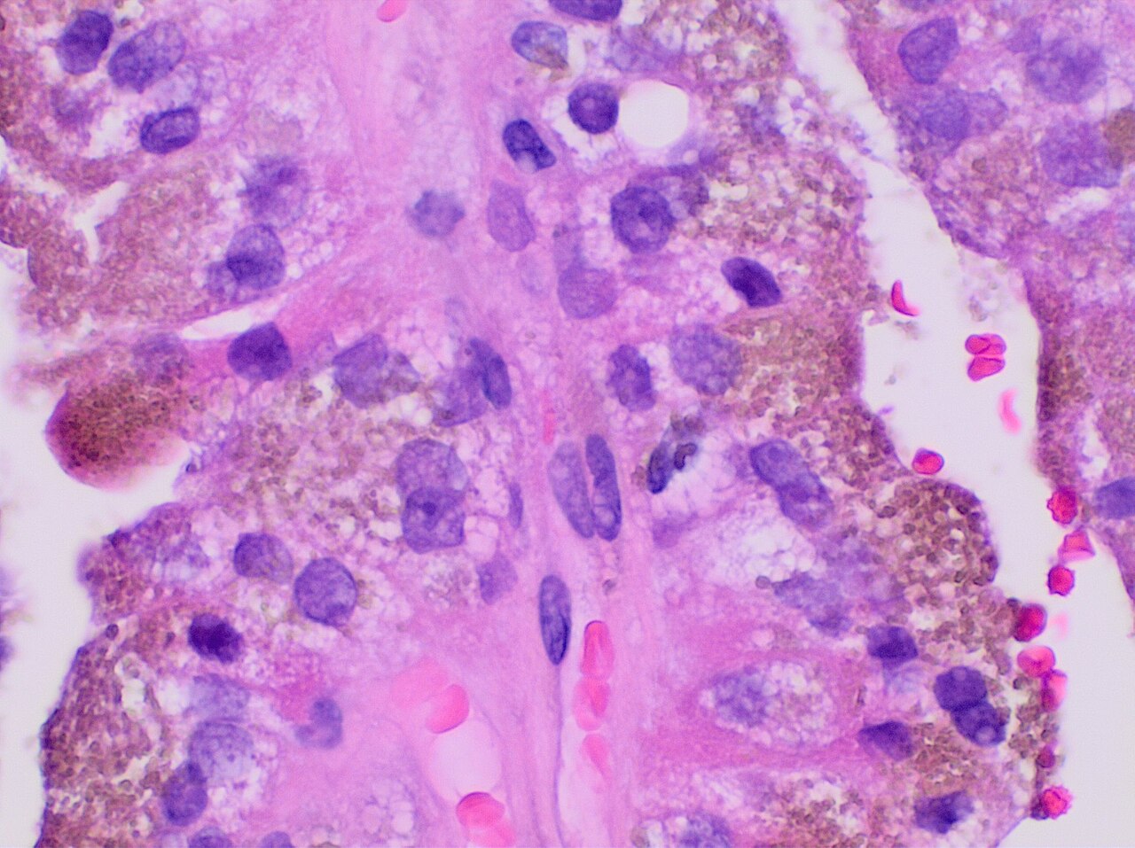
Micrograph of renal cell carcinoma in hereditary leiomyomatoCC BY-SA 3.0

Micrograph of renal cell carcinoma in hereditary leiomyomatoCC BY-SA 3.0

Micrograph of renal cell carcinoma in hereditary leiomyomatoCC BY-SA 3.0

Micrograph of renal cell carcinoma in hereditary leiomyomatoCC BY-SA 3.0

Micrograph of renal cell carcinoma in hereditary leiomyomatoCC BY-SA 3.0

Micrograph of renal cell carcinoma in hereditary leiomyomatoCC BY-SA 3.0
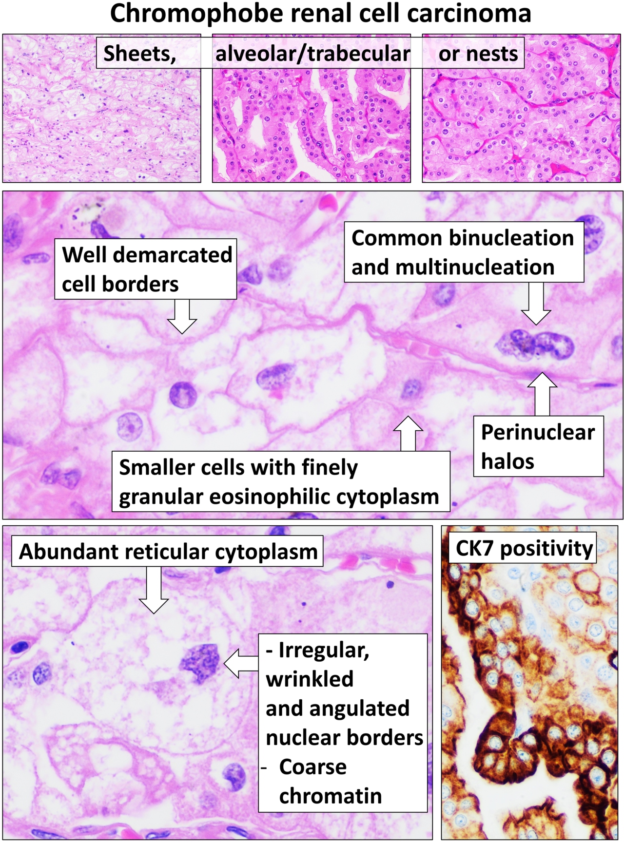
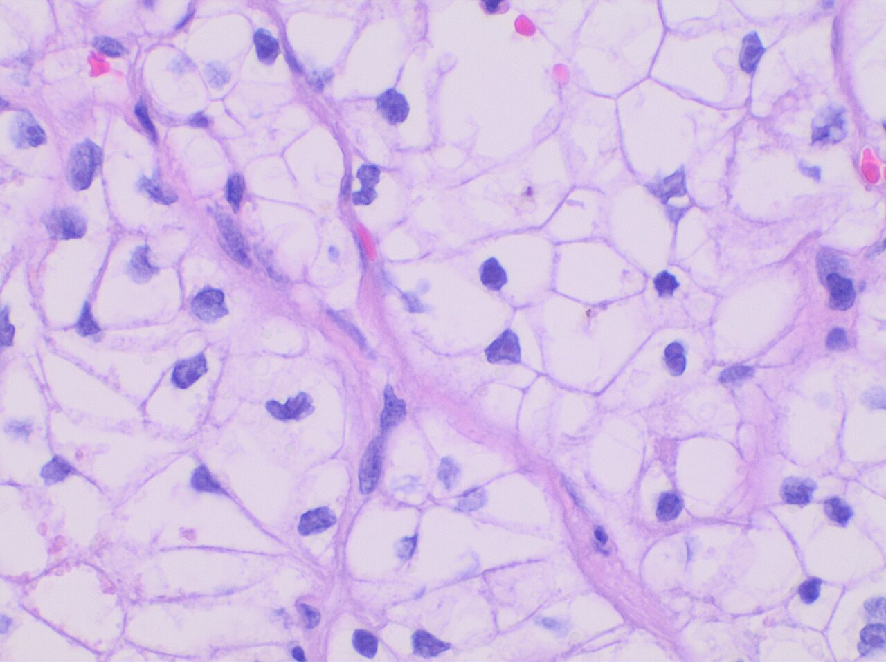
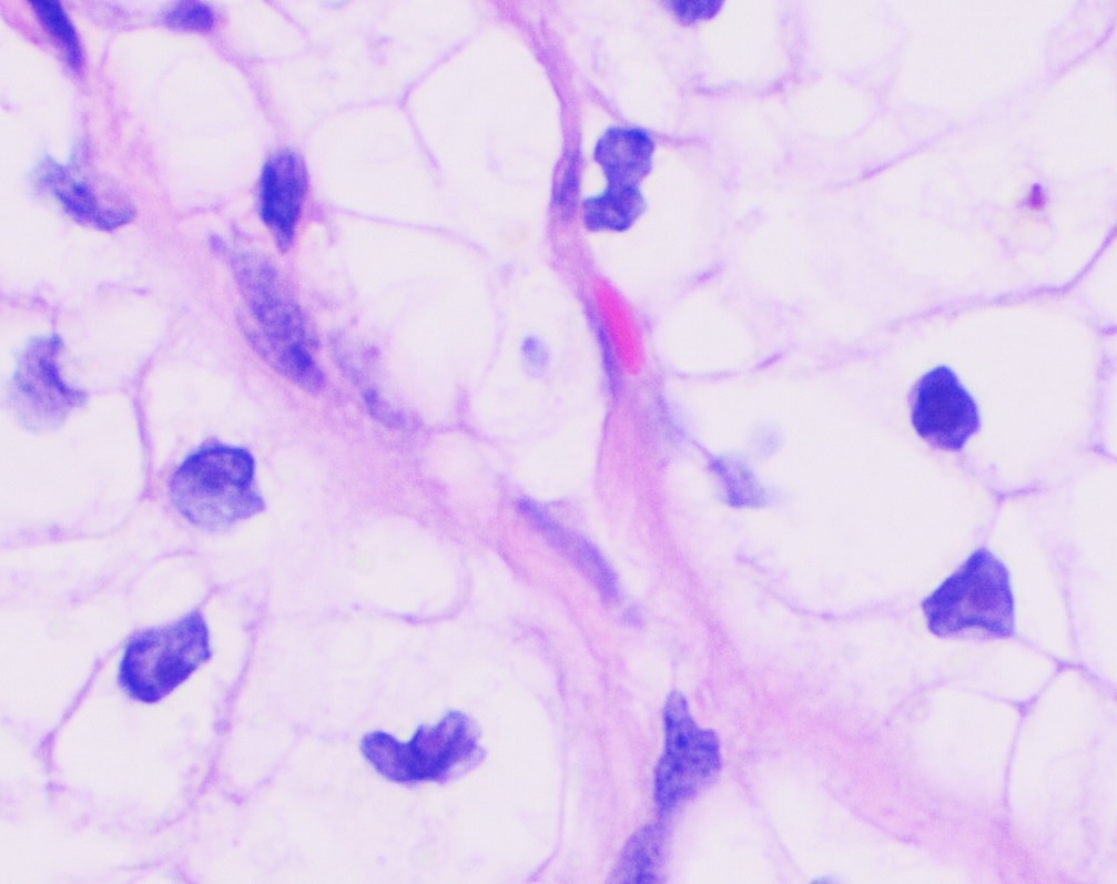
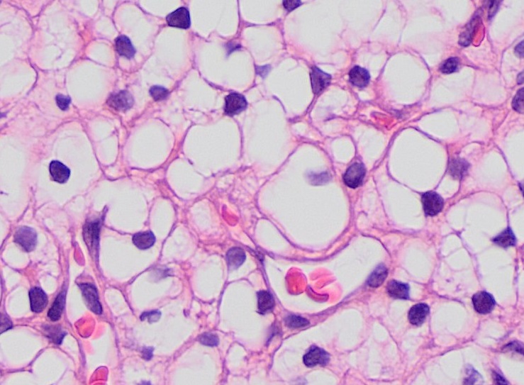
Histopathology of chromophobe renal cell carcinoma h amp e s

Histopathology of classic chromophobe renal cell carcinoma wCC BY 4.0

Histopathology of clear cell papillary renal cell carcinomaCC BY 3.0
Histopathology of clear cell renal cell carcinoma grade 1 hi
Histopathology of clear cell renal cell carcinoma grade 1 in
Histopathology of clear cell renal cell carcinoma grade 1 lo

Histopathology of eosinophilic chromophobe renal cell carcinCC BY 4.0
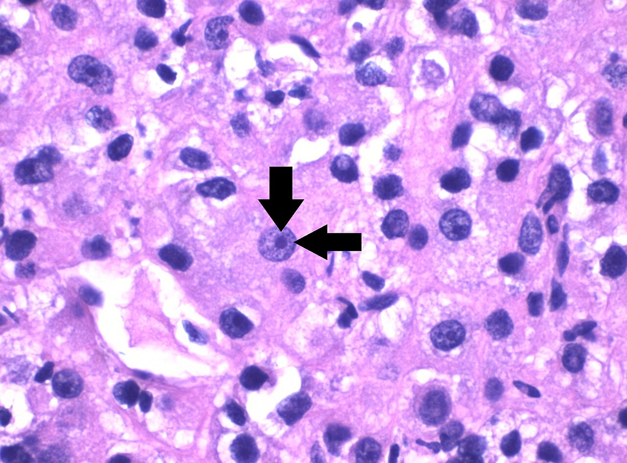
Histopathology of grade 3 clear cell renal cell carcinoma di
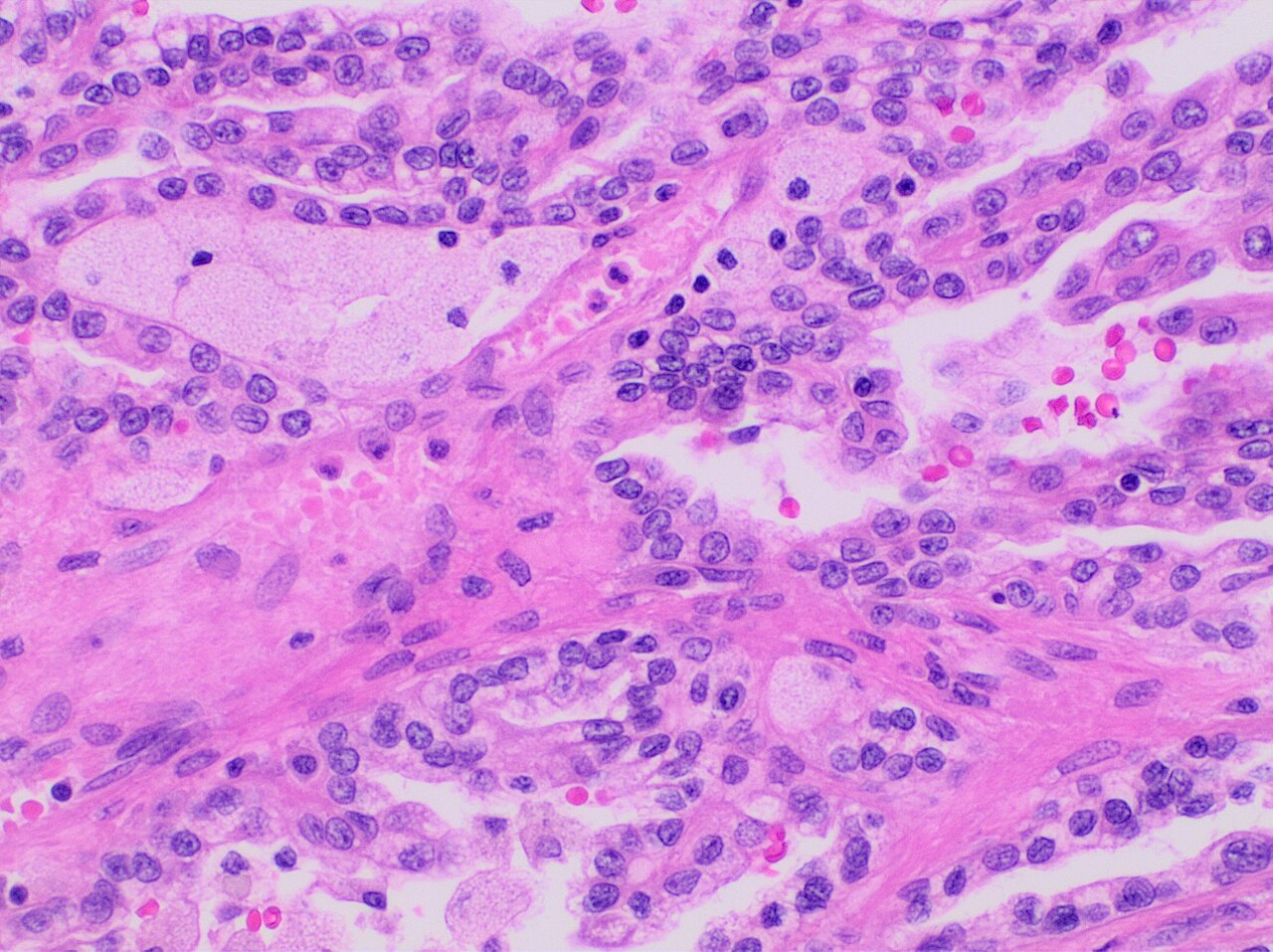
Histopathology of papillary renal cell carcinoma type 1 high
Histopathology of papillary renal cell carcinoma type 1 low

Histopathology of papillary renal cell carcinoma type 1 charCC BY 4.0

Histopathology of papillary renal cell carcinoma type 2CC BY 4.0
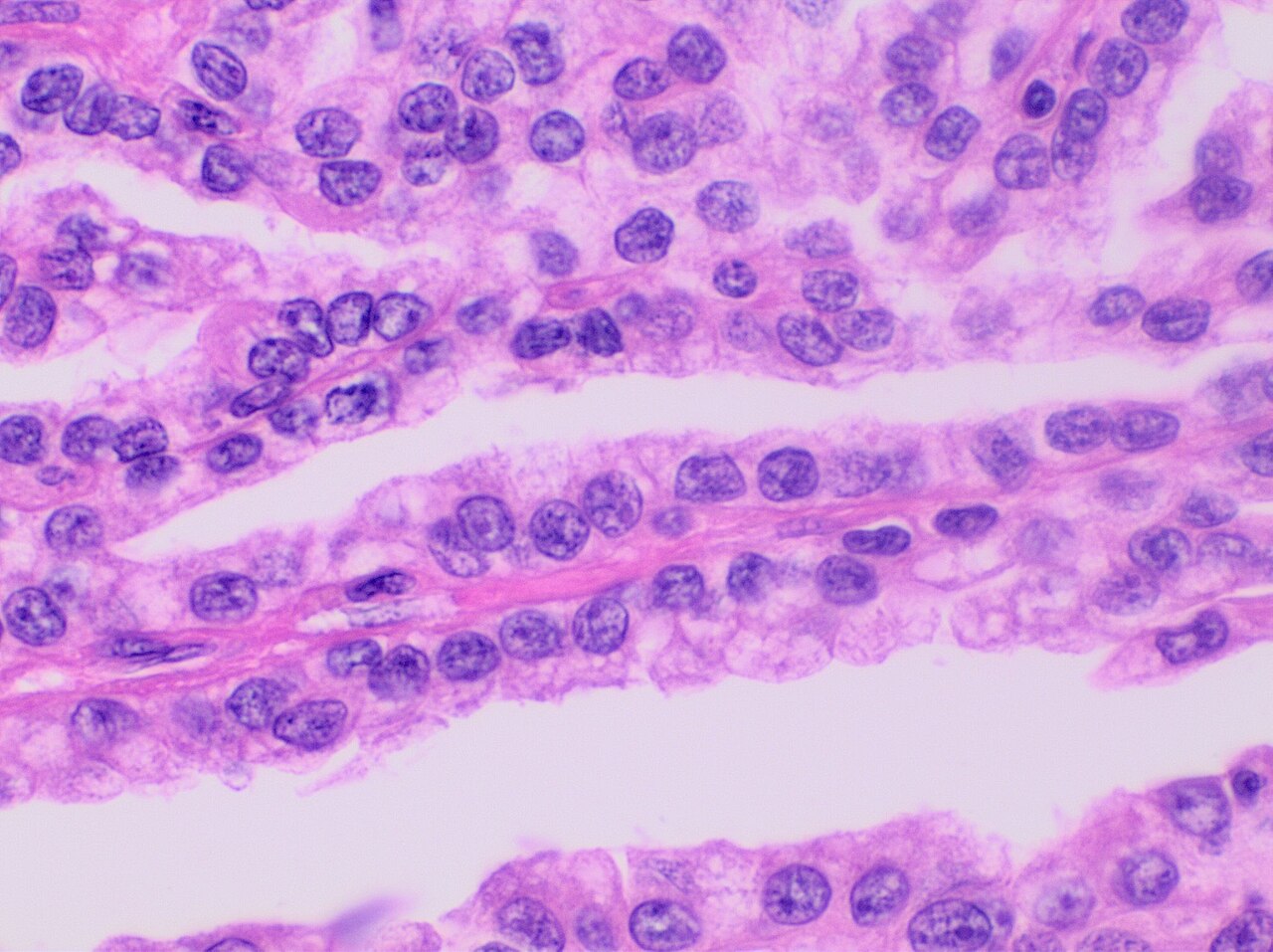
Histopathology of papillary renal cell carcinoma renal cell

Micrograph of papillary renal cell carcinoma abbreviated parCC BY-SA 3.0

Renal clear cell ca 1 nephrectomyCC BY-SA 3.0

Micrograph of an unclassified renal cell carcinoma abbreviatCC BY-SA 3.0
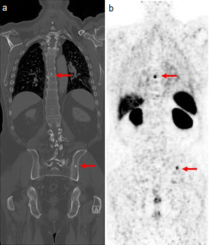
68ga psma ligand pet ct in a 78 year old patient with primar Maurer, Tobias

Metastatic renal cell carcinoma ct scan case 262CC BY-SA 2.0

Fdg pet ct scan of patient shown in image renalcancer 700mbqCC0

Fdg pet ct scan of a bone metastasis of kidney cancer the fdCC0

Sodium fluoride pet scan of a patient suffering from kidneyCC0

Sodium fluoride pet ct scan of a bone metastasis of kidney cCC0

Identifier urinaryanalysi00heit find matches title urinary aNo restrictions

Title a text book of veterinary pathology for students and pNo restrictions

Adrenal gland large adrenal cortical carcinoma this large adPublic domain

Lower respiratory tract diffuse pleural metastasis simulatinPublic domain

Identifier fibroidsalliedtu00lockuoft find matches title fibNo restrictions

Identifier fibroidsalliedtu00lockuoft find matches title fibNo restrictions
Cystoma of kidney identifier cu31924104226240 find matches tInternet Archive Book Images
Identifier cu31924104226240 find matches title general pathoInternet Archive Book Images

Left renal tumor with inferior vena cava thrombus into the rCC BY-SA 4.0

Malignant kidney tumour in a 66 year old man with primary faCC BY 4.0

Identifier manualofpatholog00coplrich find matches title manNo restrictions

Identifier oralpathologyp00barr find matches title oral pathNo restrictions

Vaginal prolapse identifier pathologytreatme00mart find matcNo restrictions

Progressive migrating tumours presented as swollen manifestaCC BY 4.0

Id 863 description gross pathology of bisected kidney showinPublic domain

Kidney bladder urinary rhabdoid tumor characteristic appearaPublic domain

Fig 204 the kidney tumor itself after removal in the case prNo restrictions

Identifier pathologysurgica1895senn find matches title the pNo restrictions

Fig 91 adenoma of the kidney after edmunds identifier patholNo restrictions

Identifier pathologysurgica1895senn find matches title the pNo restrictions

Identifier transactionsofam2319amer find matches title transNo restrictions

Kidney bladder urinary nephroblastoma note the prominent sepPublic domain

Nephroblastoma humanCC BY-SA 4.0

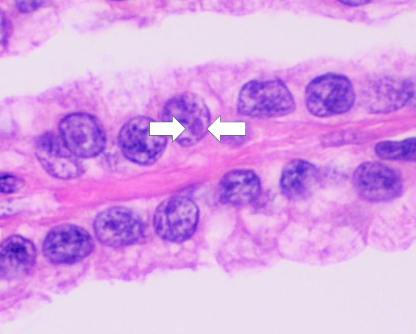
Triphasic pattern showing tubules solid sheets of cells andCC BY 2.0

Tubular pattern resembling pseudo rosettes at placesCC BY 2.0

Identifier annalsofsurgery78philuoft find matches title annaNo restrictions

Identifier annalsofsurgery78philuoft find matches title annaNo restrictions

Identifier gynecologygrav2 find matches title gynecology yeaNo restrictions

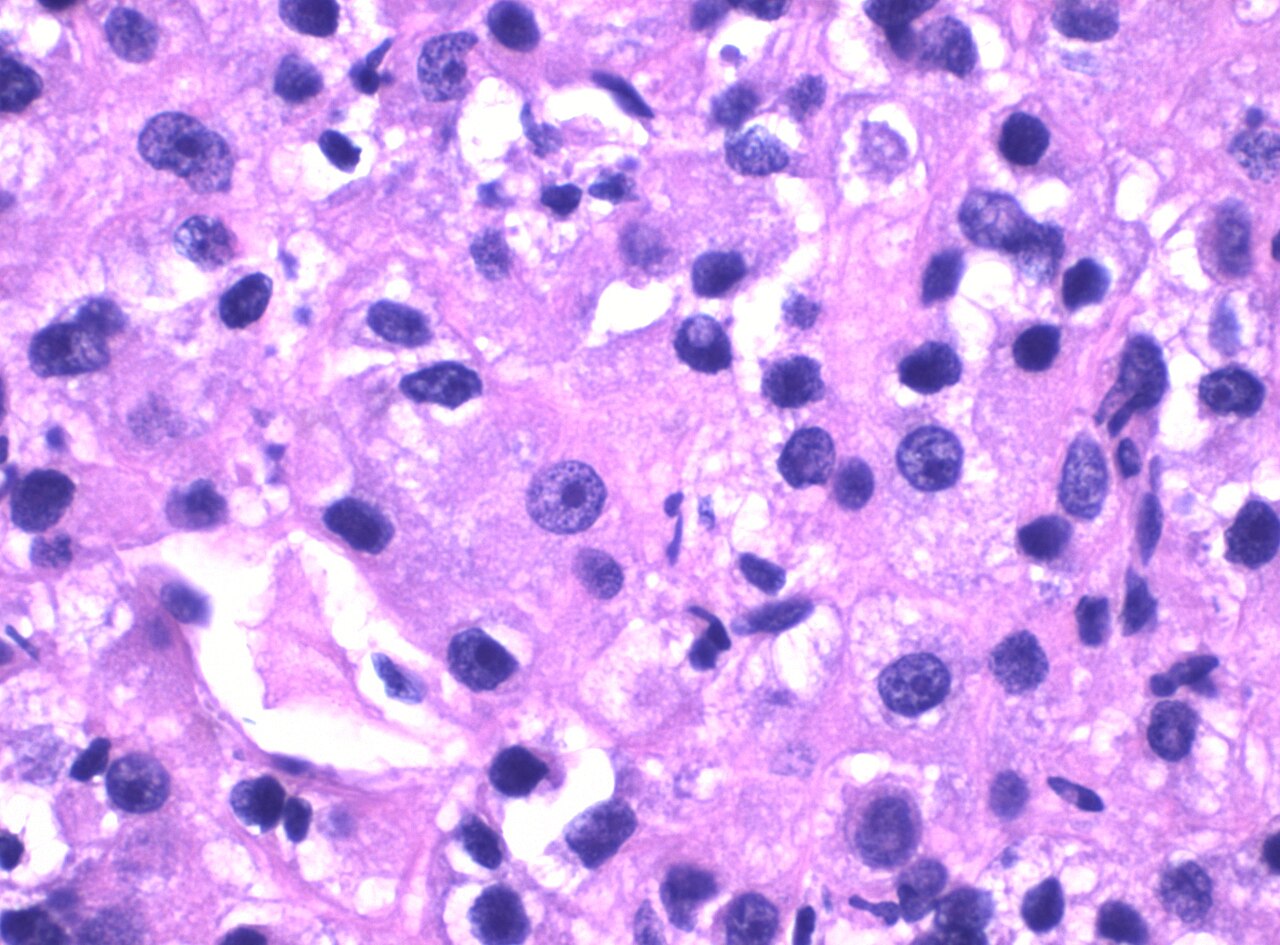
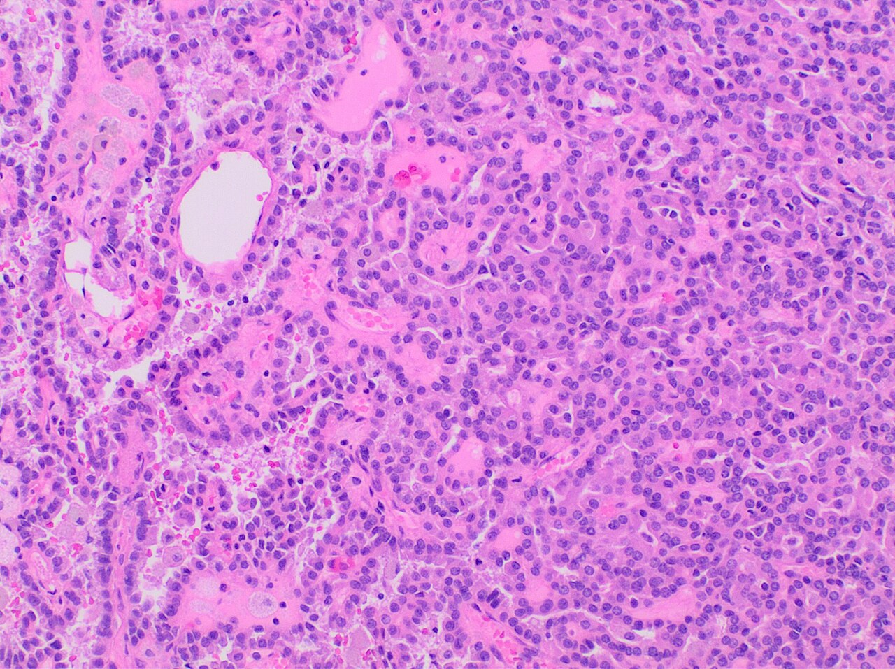
Histopathology of wilms tumor showing its three elements bla
Histopathology of wilms tumor tumor showing its three elemen

Low magnification micrograph of a wilms tumour infiltrating

22 month old girl trying out hats to wear after chemotherapyPublic domain
Whole slide image of wilms tumor h amp e stain the whole sli
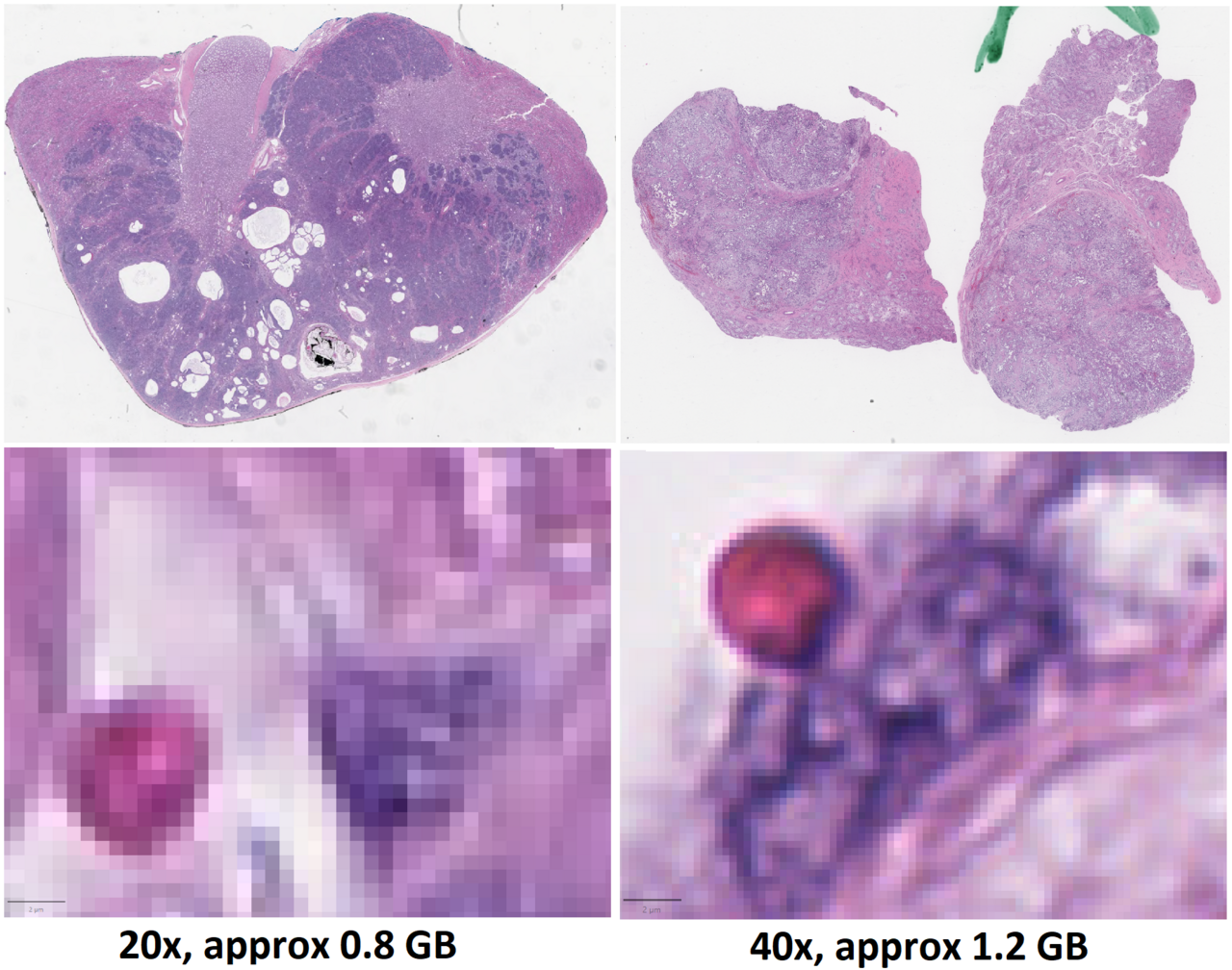
Whole slide image quality comparison images to the left are
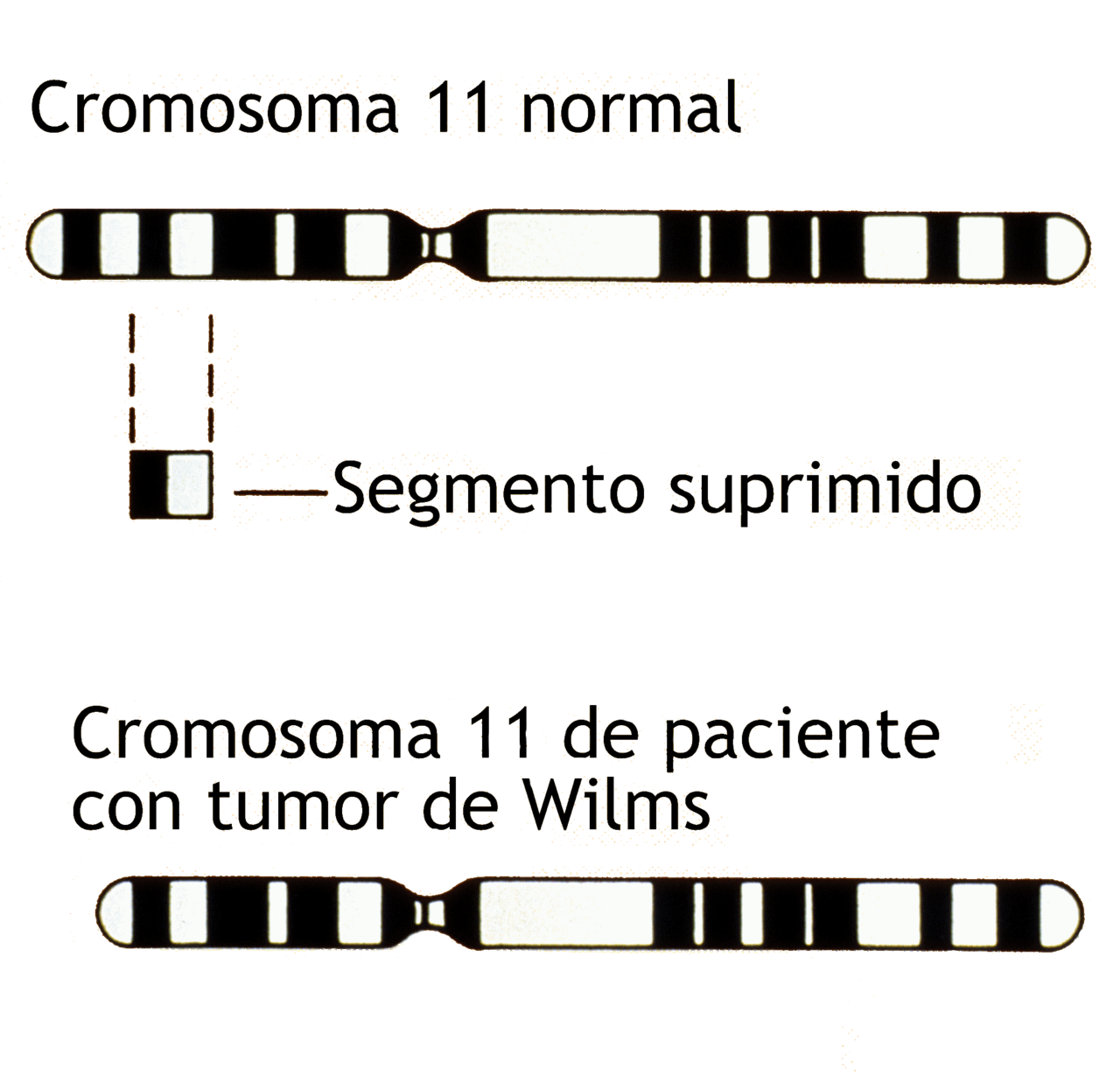
Ilustraci n que compara un cromosoma 11 normal y un cromosom

Title wilms tumor illustration description an illustration cPublic domain

WilmsPublic domain

Wilm s tumor nephroblastoma pathological and histological imCC BY 2.0

Ct scan of 11 cm wilms tumor nephroblastoma of right kidneyPublic domain

A tumor is seen in the upper pole of the kidney the tumor isCC BY-SA 4.0

Wt 1 immunohistochemistry cytoplasmatic staining in glioblasCC BY-SA 4.0

High magnification micrograph of a wilms tumour also nephrobCC BY-SA 3.0

Intermediate magnification micrograph of a wilms tumour alsoCC BY-SA 3.0

Low magnification micrograph of a wilms tumour also nephroblCC BY-SA 3.0

Very high magnification micrograph of a wilms tumour also neCC BY-SA 3.0

Micrograph showing metastatic clear cell renal cell carcinomCC BY-SA 4.0

Micrograph showing metastatic clear cell renal cell carcinomCC BY-SA 4.0

Micrograph showing metastatic clear cell renal cell carcinomCC BY-SA 4.0
Histopathology of grade 3 clear cell renal cell carcinoma di
Histopathology of papillary renal cell carcinoma type 1 grad
Histopathology of papillary renal cell carcinoma type 1 grad
Histopathology of papillary renal cell carcinoma type 1 grad

Histopathology of conventional renal clear cell carcinomaCC BY 4.0

Adult polycystic kidney from a bilateral nephrectomy specimeCC BY 2.0

Diffuse proliferative lupus nephritis this photo of type ivPublic domain

Gross appearance of the cut surface of a nephrectomy specimeCC BY 3.0

Histopathologic image of renal angiomyolipoma nephrectomy spCC BY-SA 3.0

Histopathologic image of renal angiomyolipoma nephrectomy spCC BY-SA 3.0

Histopathologic image of renal angiomyolipoma nephrectomy spCC BY-SA 3.0

Histopathologic image of renal angiomyolipoma nephrectomy spCC BY-SA 3.0

Gross appearance of the cut surface of a nephrectomy specimeCC BY 3.0

Histopathological image of renal oncocytoma nephrectomy specCC BY-SA 3.0

Identifier transactionsofso2119sout find matches title transNo restrictions

Renal cell carcinoma with both cystic and solid components lCC BY 4.0

Solid tumor in the renal sinus seen as a hypoechoic mass latCC BY 4.0

Contrast enhanced ct of the abdomen of a 75 year old woman pCC0
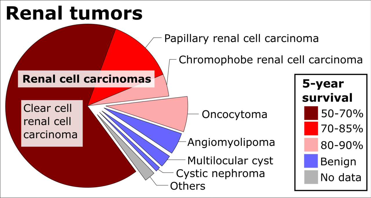
Edit renal tumors by relative incidence and prognosis refere
Edit renal tumors by relative incidence and prognosis refere
Cancerologie - vessie
98 images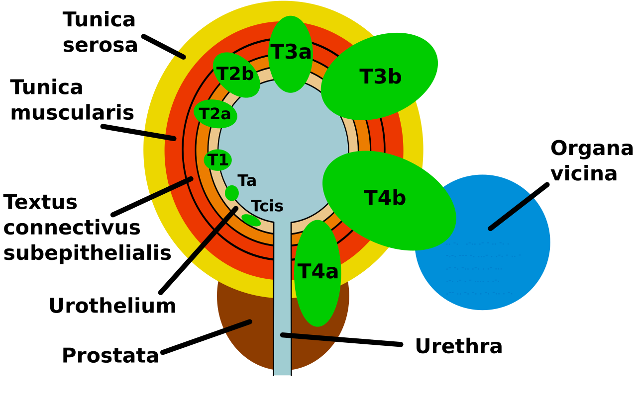
Bladder cancer tnm classification

3d medical animation still showing urinary bladder cancerCC BY-SA 4.0

Bladder cancer treatment flowchartCC BY-SA 3.0

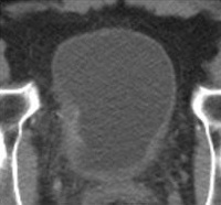
Bladder wall thickening due to cancer this is an edited versCC BY-SA 4.0

Transitional cell carcinoma of the bladder the white in theCC BY-SA 4.0

This svg file was uploaded with commonist disability adjusteCC BY-SA 4.0

This svg file was uploaded with commonist disability adjusteCC BY-SA 4.0

Age standardised death rates from bladder cancer by countryCC BY-SA 2.5

Micrograph showing invasion of the urinary bladder neck by pCC BY-SA 4.0

Histopathology of urothelial carcinoma of the urinary bladdeCC BY-SA 3.0

Bladder cancer see a full animation of this medical topicCC BY 3.0
Ct scan of a bladder cancer Ravi K. Samala

Cancer of the gall bladder wellcome images keywords neoplasmCC BY 4.0

Cancer prostate extending into bladder walll with chronic inCC0

Diagram showing advanced bladder cancerCC BY-SA 4.0

Diagram showing early stage bladder cancerCC BY-SA 4.0

Diagram showing invasive bladder cancerCC BY-SA 4.0

Diagram showing stage n1 bladder cancerCC BY-SA 4.0

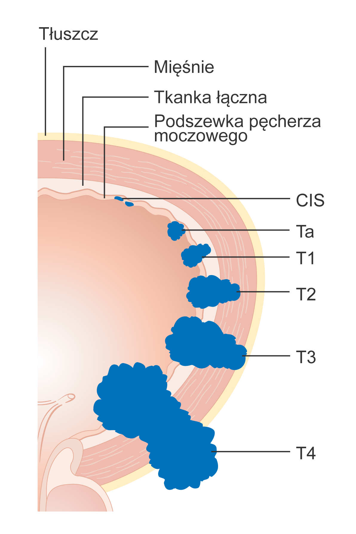
Diagram showing the t stages of bladder cancerCC BY-SA 4.0
Schemat zaawansowania miejscowego raka p cherza moczowego
Histopathology of keratinizing squamous cell carcinoma of th

Histopathology of keratinizing squamous cell carcinoma of th

Gross pathology of papillary urothelial carcinoma of bladderCC BY-SA 3.0

A 3d render of the urinary bladder of a human maleCC BY-SA 4.0

T stages of bladder cancer the white in the bladder is contrCC BY-SA 4.0

Clear cell papillary urothelial carcinoma h amp e stainCC0

Clear cell papillary urothelial carcinoma h amp e stainCC0

Clear cell papillary urothelial carcinoma h amp e stainCC0

Clear cell urothelial carcinoma displaying cells with abundaCC0

Clear cell urothelial carcinoma displaying cells with abundaCC0

Clear cell urothelial carcinoma displaying cells with abundaCC0

Clear cell urothelial carcinoma displaying cells with abundaCC0

Micropapillary urothelial carcinoma characterized by micropaCC0

Micropapillary urothelial carcinoma characterized by micropaCC0

Micropapillary urothelial carcinoma characterized by micropaCC0

Micropapillary urothelial carcinoma characterized by micropaCC0

High grade noninvasive papillary urothelial carcinoma characCC0

Papillary urothelial carcinoma showing a cribriform morpholoCC0

Papillary urothelial carcinoma showing a cribriform morpholoCC0

Plasmacytoid urothelial carcinoma showing monotonous discoheCC0

Plasmacytoid urothelial carcinoma showing monotonous discoheCC0

Plasmacytoid urothelial carcinoma with lymphovascular invasiCC0

Stage pt1 urothelial carcinoma with inflammatory stromal reaCC0

Urothelial carcinoma with glandular differentiation showingCC0

Urothelial carcinoma with glandular differentiation showingCC0

Invasive urothelial carcinoma showing giant bizarre anaplastCC0

Micropapillary urothelial carcinoma in a resection specimenCC0

Micropapillary urothelial carcinoma in a resection specimenCC0

Urothelial carcinoma with squamous differentiation h amp e sCC0

Urothelial carcinoma showing glandular differentiation and vCC0

Identifier manualofmodernsu1899robe find matches title a manNo restrictions

A blue light cystoscopy includes use of an imaging agent thaCC BY-SA 4.0

Blue light cystoscopy performed in 2007 or 2008 at sf ioan cCC BY 4.0

Figure 2 cystoscopy image shows a calculus connected to theCC BY 4.0

Identifier modernsurgerygen1919daco find matches title moderNo restrictions

Identifier operativegynecol001kell find matches title operatNo restrictions

Identifier surgeryitsprinci04keen find matches title surgeryNo restrictions

Micrograph showing metastatic clear cell renal cell carcinomCC BY-SA 4.0

Micrograph of granulomatous inflammation of the urinary bladCC BY-SA 3.0

High magnification micrograph of granulomatous inflammationCC BY-SA 3.0

H amp e representative low power view of the tumor without tCC BY-SA 2.0

Sarcomatoid carcinoma of the urinary bladder tur specimen hCC0

Sarcomatoid carcinoma of the urinary bladder featuring fasciCC0

Sarcomatoid carcinoma of the urinary bladder tur specimen hCC0

Sarcomatoid carcinoma of the urinary bladder tur specimen hCC0

Sarcomatoid carcinoma of the urinary bladder featuring fasciCC0

Sarcomatoid carcinoma of the urinary bladder tur specimen hCC0

Sarcomatoid carcinoma of the urinary bladder featuring fasciCC0

Sarcomatoid carcinoma of the urinary bladder tur specimen hCC0

Sarcomatoid carcinoma of the urinary bladder featuring fasciCC0

Micrograph of small cell carcinoma of the urinary bladder hCC BY-SA 3.0

Micrograph of small cell carcinoma of the urinary bladder hCC BY-SA 3.0

Micrograph of small cell carcinoma of the urinary bladder hCC BY-SA 3.0

Urothelial carcinoma nested variant showing deceptively beniCC0

Urothelial carcinoma nested variant showing deceptively beniCC0

Urothelial carcinoma nested variant showing deceptively beniCC0

Clear cell urothelial carcinoma positive gata3 immunohistochCC0

Clear cell urothelial carcinoma positive gata3 immunohistochCC0

Micrograph showing micropapillary urothelial carcinoma alsoCC BY-SA 3.0

Micrograph showing micropapillary urothelial carcinoma alsoCC BY-SA 3.0

Micrograph showing micropapillary urothelial carcinoma alsoCC BY-SA 3.0

Micrograph showing micropapillary urothelial carcinoma alsoCC BY-SA 3.0

Micrograph showing micropapillary urothelial carcinoma alsoCC BY-SA 3.0

High magnification micrograph of nested variant of urotheliaCC BY-SA 3.0

Micrograph of plasmacytoid urothelial carcinoma also plasmacCC BY-SA 3.0

Micrograph of plasmacytoid urothelial carcinoma also plasmacCC BY-SA 3.0

Micrograph of plasmacytoid urothelial carcinoma also plasmacCC BY-SA 3.0

Micrograph of plasmacytoid urothelial carcinoma also plasmacCC BY-SA 3.0

Micrograph of plasmacytoid urothelial carcinoma also plasmacCC BY-SA 3.0

Micrograph of plasmacytoid urothelial carcinoma also plasmacCC BY-SA 3.0

Micrograph of plasmacytoid urothelial carcinoma also plasmacCC BY-SA 3.0

Micrograph of urothelial cell carcinoma of the prostatic ureCC BY-SA 3.0

Micrograph of urothelial carcinoma also urothelial cell carcCC BY-SA 3.0

Micrograph of urothelial carcinoma also urothelial cell carcCC BY-SA 3.0

Bladder tumor in fdg pet ct suv 10 5 due to the high physiolPublic domain

Identifier urinaryanalysi00heit find matches title urinary aNo restrictions

Identifier urinaryanalysi00heit find matches title urinary aNo restrictions

Identifier nursinginabdomi00full find matches title nursingNo restrictions
Cancerologie - prostate
72 images
Anterior x ray view of prostate cancerCC BY-SA 4.0

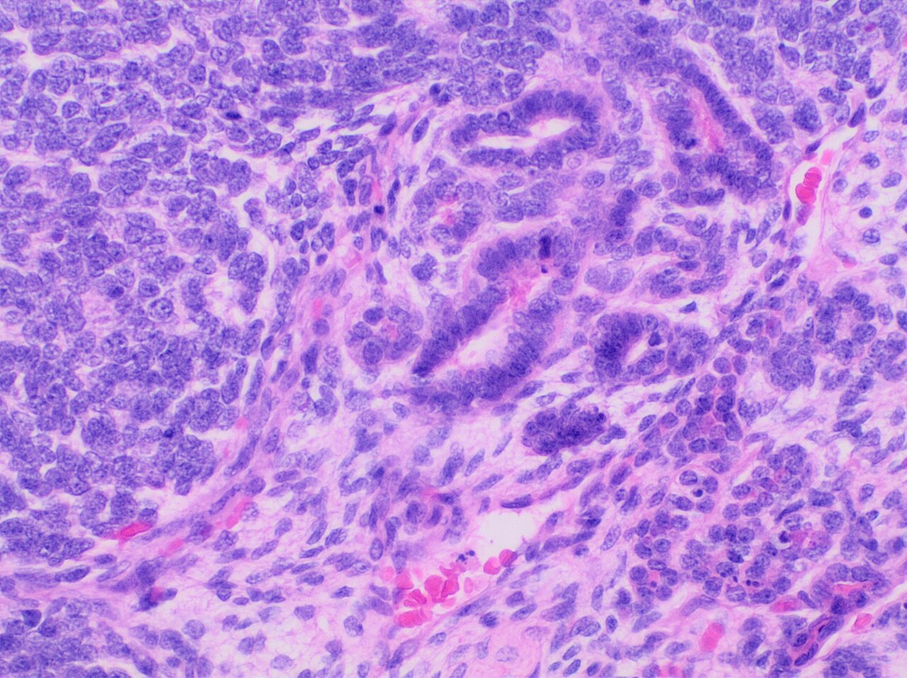
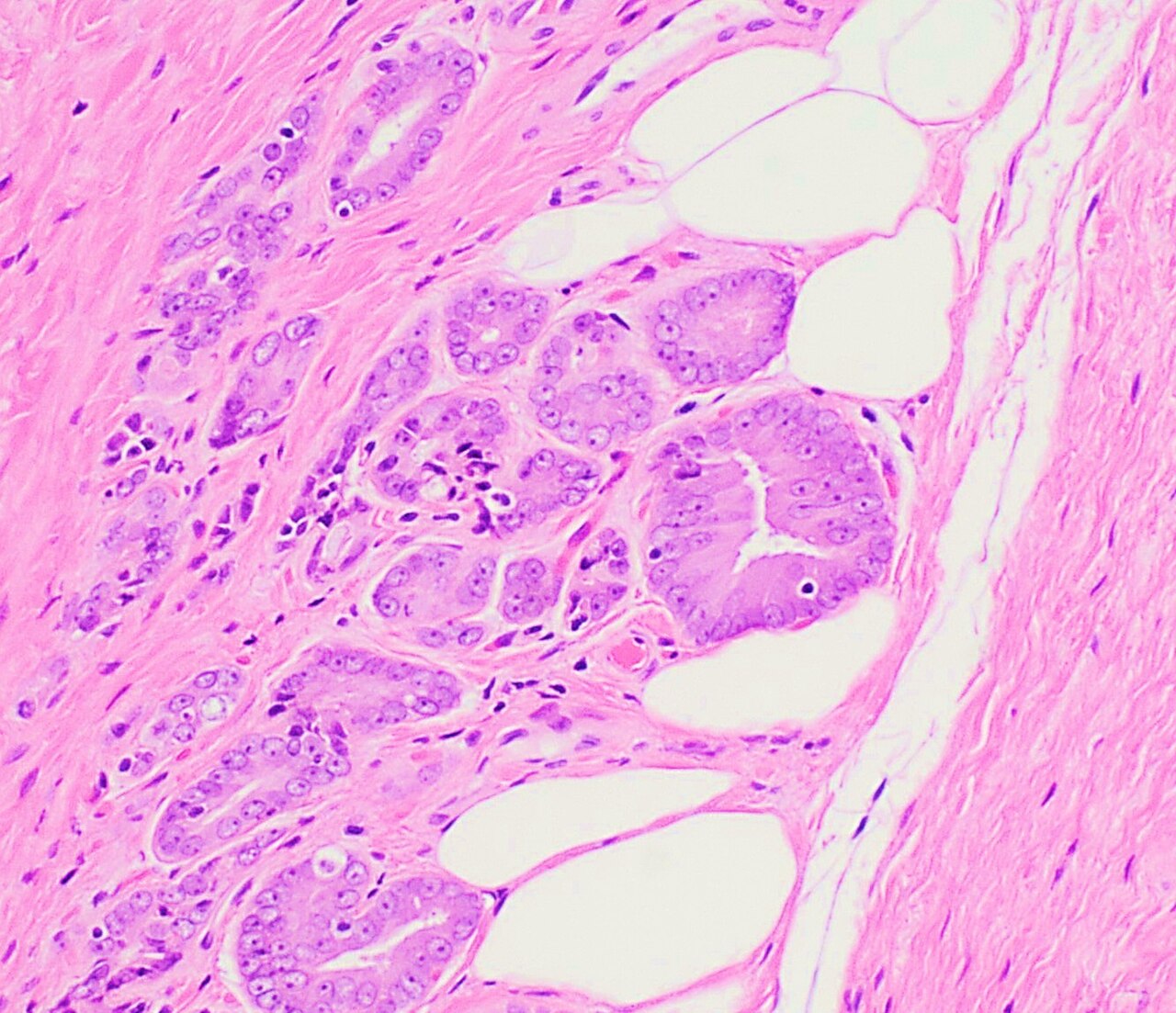
Adenocarcinoma of the prostate on the photomicrograph you caCC BY-SA 4.0

Diagnosis of prostate cancerCC BY 4.0

Diagram showing t1 3 stages of prostate cancerCC BY-SA 4.0

Biopsja gruczo u krokowegoCC BY-SA 4.0

Diagram showing prostate cancer pressing on the urethraCC BY-SA 4.0

Diagram showing stage t4 prostate cancerCC BY-SA 4.0

Human prostate cancer cells undergoing ferroptosis capturedCC BY-SA 4.0
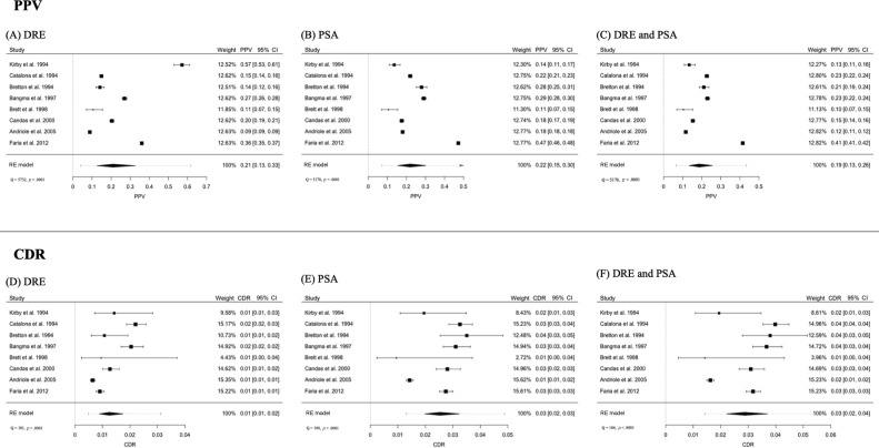
Forest plots for the ppv of a dre b psa and c dre psa and th

Infographic on prostate cancer and its statistics in franceCC BY-SA 4.0

Illustrative images of some morphological criteria for the dCC BY 4.0

Differences between healthy prostate and a prostate with a tCC BY-SA 3.0

Prostate cancer see a full animation of this medical topicCC BY-SA 4.0

New findings on prostate cancer may enable doctors to make bCC BY 2.0

Table outlining risk stratification for prostate cancerCC BY-SA 3.0

Title prostate cancer description histological slide h amp ePublic domain

Table of comparison of prostate screening results globallyPublic domain

The ohio cancer incidence surveillance system ociss ohio depPublic domain

Age standardised death rates from prostate cancer by countryCC BY-SA 2.5

Ratio of annual mortality rates in east and west germany forCC BY 4.0

A lateral x ray view of prostate cancerCC BY-SA 4.0

Anterior x ray view of prostate cancerCC BY-SA 4.0

Zero the end of prostate cancer logoPublic domain

Micrograph showing ductal adenocarcinoma of the prostate glaCC BY-SA 3.0

Micrograph showing ductal adenocarcinoma of the prostate glaCC BY-SA 3.0

Micrograph showing ductal adenocarcinoma of the prostate glaCC BY-SA 3.0

Micrograph showing ductal adenocarcinoma of the prostate glaCC BY-SA 3.0
Histopathology of prostate adenocarcinoma involving adipose
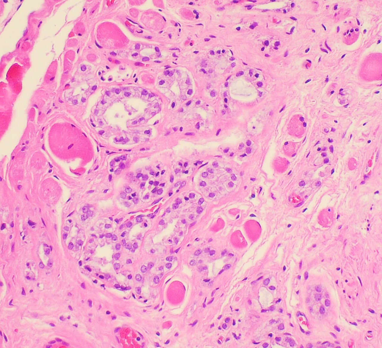
Histopathology of prostate adenocarcinoma involving skeletal
Histopathology of prostatic adenocarcinoma gleason 3 3 6 wit

Micrograph of prostate adenocarcinoma with a glomeruloid glaCC BY 4.0

Micrograph showing mucinous adenocarcinoma of the prostate hCC BY-SA 4.0

Micrograph showing mucinous adenocarcinoma of the prostate hCC BY-SA 4.0

Micrograph showing mucinous adenocarcinoma of the prostate hCC BY-SA 4.0

Histopathologic image of acinar type adenocarcinoma obtainedCC BY-SA 3.0

Histopathologic image of acinar type adenocarcinoma obtainedCC BY-SA 3.0

Histopathologic image of acinar type adenocarcinoma obtainedCC BY-SA 3.0

Histopathologic image of acinar type adenocarcinoma obtainedCC BY-SA 3.0

Histopathologic image of acinar type adenocarcinoma obtainedCC BY-SA 3.0

Prostate adenocarcinoma 6CC BY-SA 3.0
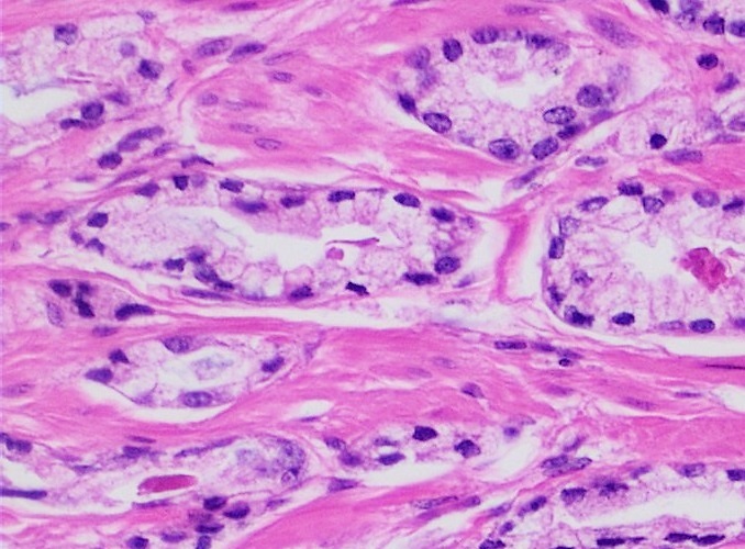
Prostate adenocarcinoma acinar pattern h amp e stain

Microsopic view hematoxilin amp eosin 100x of abnormal tissuCC BY-SA 4.0

Intermediate magnification micrograph of prostate adenocarciCC BY-SA 3.0

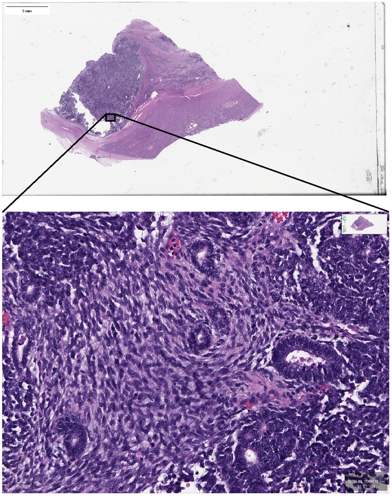
Micrograph of prostate adenocarcinoma needle core biopsy hpsCC BY-SA 3.0

Whole slide of half a prostate scanned epson perfection 1670CC BY-SA 3.0
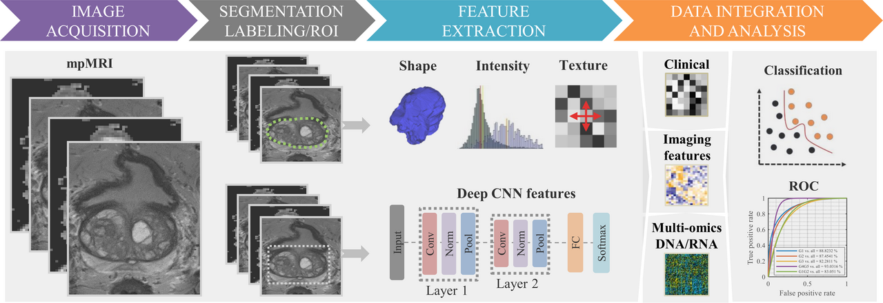
Flowchart of the standard radiomics model 1 multiparametric Kucharczyk, M.J.

Mri showing hydrogel spacer pushing the rectum away from theCC BY-SA 3.0

Diagram showing a prostate biopsyCC BY-SA 4.0

Esquema que mostra una bi psia transperineal de pr stataCC BY-SA 3.0

Diagram showing a transperineal prostate biopsyCC BY-SA 4.0

Leonard s marks m d diagram of targeted prostate biopsy andCC BY-SA 4.0

Micrograph of acinar adenocarcinoma of the prostate with bluCC BY 4.0

Micrograph of acinar adenocarcinoma of the prostate with douCC BY 4.0

Micrograph of acinar adenocarcinoma of the prostate with mulCC BY 4.0

Micrograph of adenocarcinoma of the prostate with two mitoseCC BY 4.0

Prostate needle biopsy see a full animation of this medicalCC BY-SA 4.0

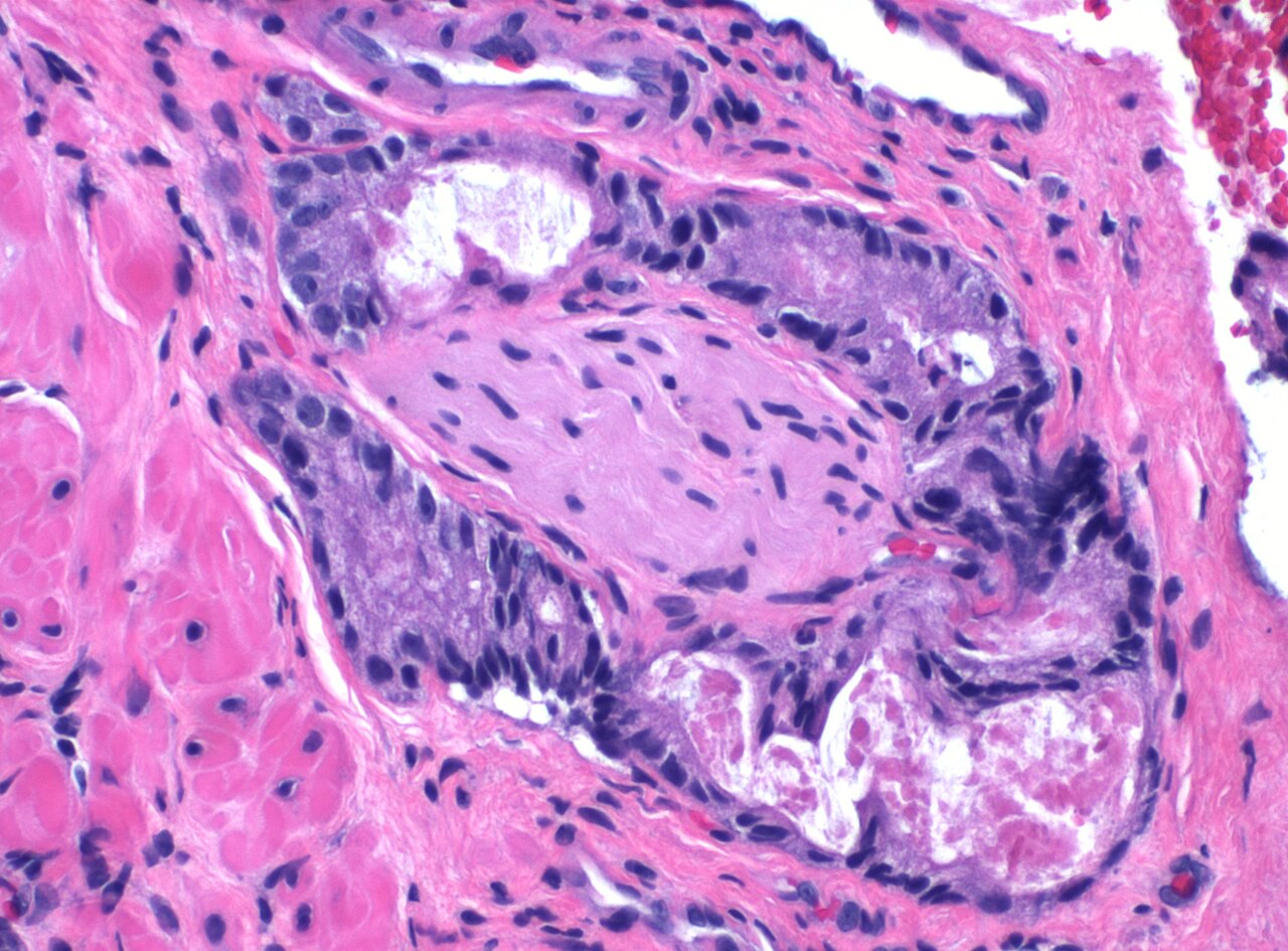
Micrograph showing a biopsy of the rectum with prostatic carCC BY-SA 3.0

Micrograph showing a biopsy of the rectum with prostatic carCC BY-SA 3.0

Micrograph showing a biopsy of the rectum with prostatic carCC BY-SA 3.0

Micrograph showing prostate carcinoma with extraprostatic exCC BY-SA 3.0

Prostate slideCC BY-SA 3.0

Targeted mri us fusion prostate biopsy at uclaCC BY-SA 3.0

GleasonscorePublic domain

Bone metastasis of prostate cancer in f 18 choline pet ct scCC BY 3.0

Osteoplastic bone metastasis of a histologically confirmed pPublic domain
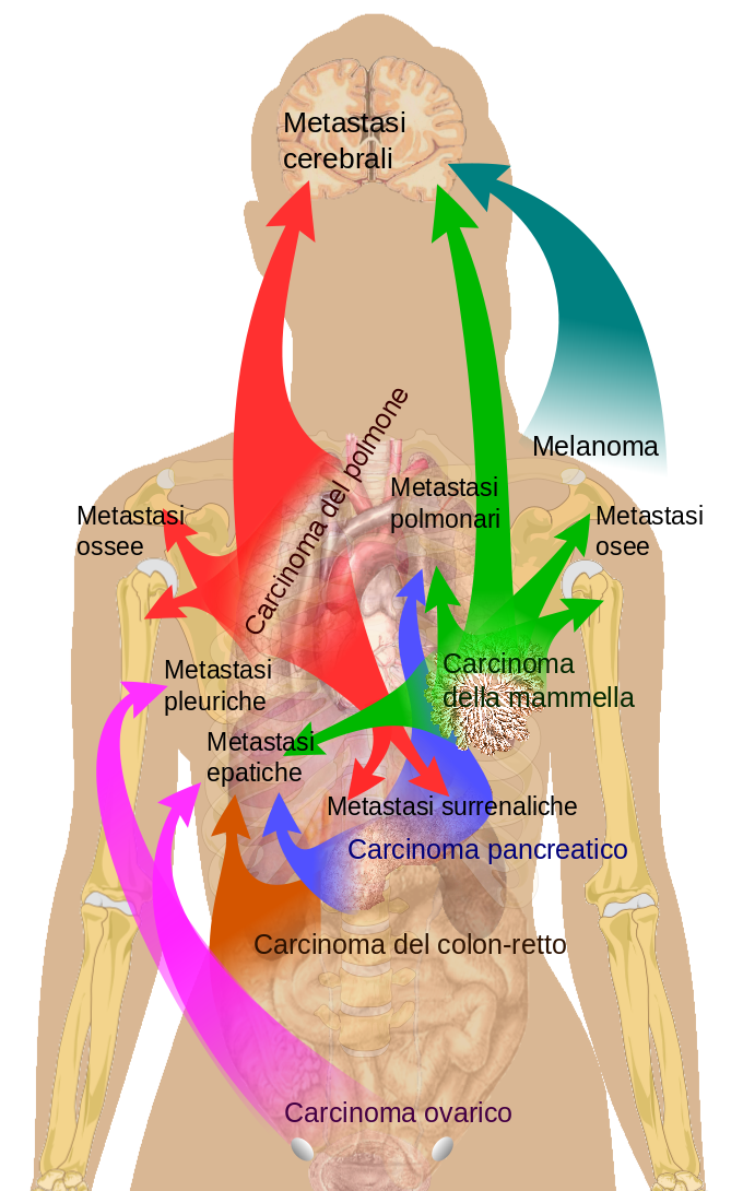
Main sites of metastases for common cancer types primary can/ref>

Main sites of metastases for common cancer types primary canCC0

Osteoplastic metastasis of prostate cancer in choline pet ctCC BY-SA 3.0

A 70 year old male having advanced prostate cancer presentinCC BY-SA 2.0
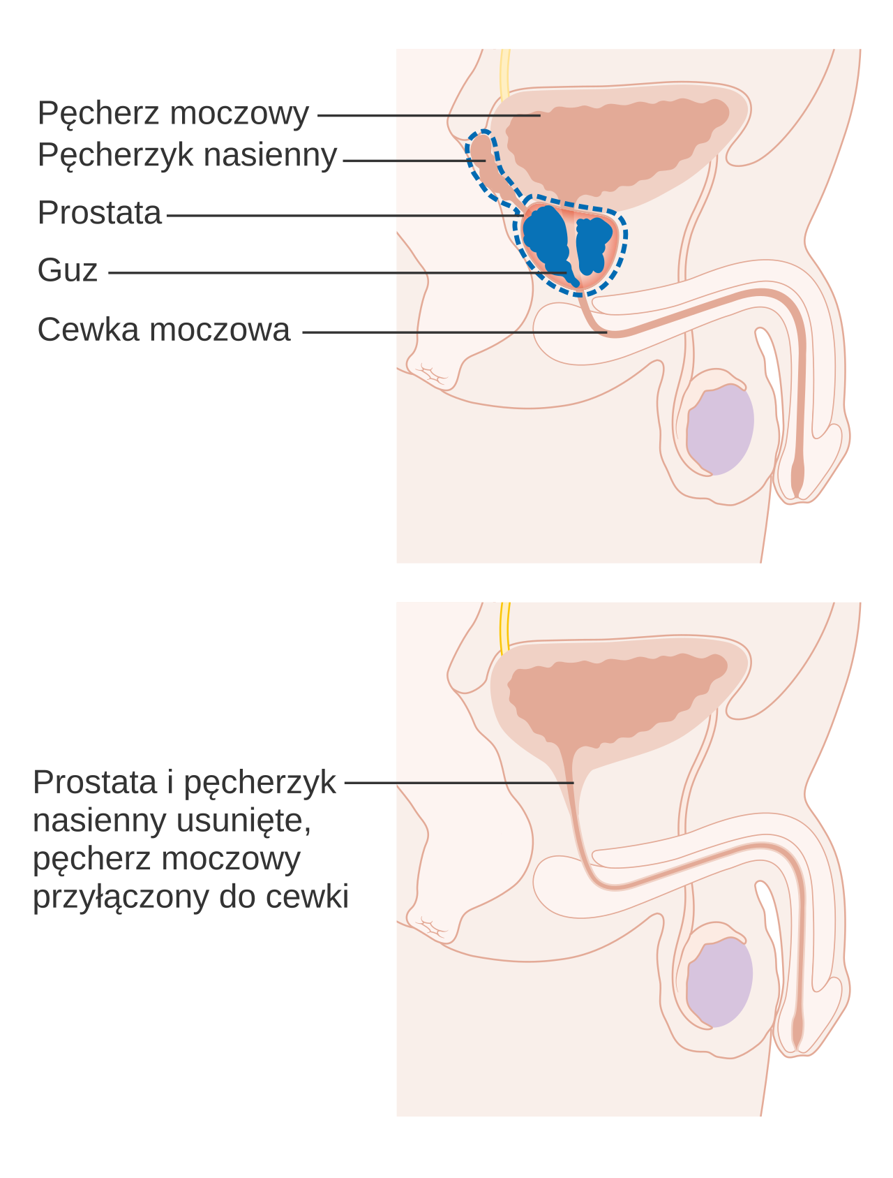
Zabieg usuni cia prostaty
Zabieg usuni cia prostaty

Diagram showing before and after a radical prostatectomyCC BY-SA 4.0
Cancerologie - testicule
63 images
Identifier annalsofsurgery78philuoft find matches title annaNo restrictions

Fertilities of men with testicular cancer compared with contCC BY-SA 3.0

Incidence standardis e pur l age pour 8 pays d europe du norCC BY-SA 3.0

Dog necropsy retained testicle with cancerCC BY 2.0

Fatal testicular cancer in a 66 year old man with secondarieCC BY 4.0

Microscopic image of testicular cancerCC BY 4.0

Photograph of a 31 year old male with a single testicle theCC BY-SA 4.0

Photograph of a 31 year old male with a single testicle theCC BY-SA 4.0

A guide on checking yourself for testicular chancer found onCC BY-SA 4.0

Founder of testicular cancer ukCC BY-SA 4.0
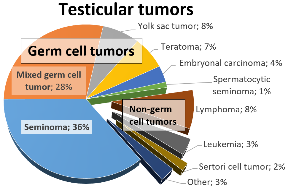
Relative incidences of testicular tumors reference gill ms s

Seminoma of the testisCC BY 2.0
Testicular cancer foundation logo 2026Public domain

September 2016Public domain

A medical illustration depicting a testicular self examinatiCC BY-SA 4.0

Lump or swelling in the testesCC BY-SA 4.0

Micrograph showing testis with metastatic prostate carcinomaCC BY-SA 3.0

Micrograph showing testis with metastatic prostate carcinomaCC BY-SA 3.0

Micrograph showing testis with metastatic prostate carcinomaCC BY-SA 3.0
Identifier tumoursinnocentm1911blan find matches title tumouInternet Archive Book Images
Histopathology of seminoma high magnification h amp e stain
Histopathology of seminoma intermediate magnification h amp
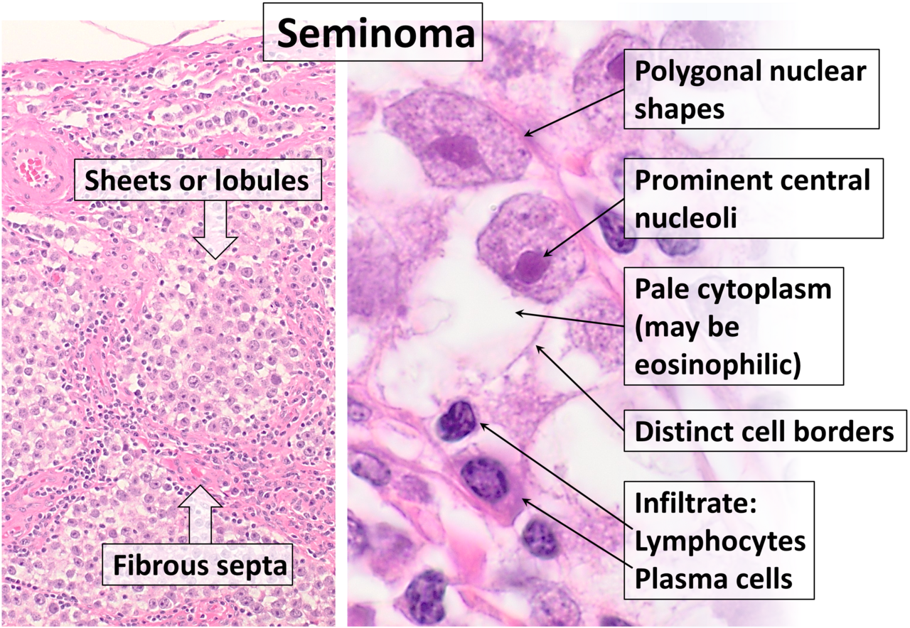
Histopathology of classical seminoma with typical features h

Micrograph showing an intertubular seminoma abbreviated itsCC BY-SA 3.0

Micrograph showing a seminoma with syncytiotrophoblasts h amCC BY-SA 4.0

Micrograph showing a seminoma with syncytiotrophoblasts h amCC BY-SA 4.0

Micrograph of a seminoma h amp e stain features lymphocytesCC BY-SA 3.0

Micrograph of a seminoma h amp e stain features lymphocytesCC BY-SA 3.0

Micrograph showing a seminoma with granulomas h amp e stainCC BY-SA 4.0

Micrograph showing a seminoma with granulomas h amp e stainCC BY-SA 4.0

Micrograph showing a seminoma with granulomas h amp e stainCC BY-SA 4.0

Micrograph showing a seminoma with granulomas h amp e stainCC BY-SA 4.0

Micrograph showing a seminoma with granulomas h amp e stainCC BY-SA 4.0

Micrograph showing a seminoma with granulomas h amp e stainCC BY-SA 4.0

Micrograph showing a seminoma with granulomas h amp e stainCC BY-SA 4.0

Micrograph showing a seminoma with granulomas h amp e stainCC BY-SA 4.0

Micrograph showing a seminoma with granulomas h amp e stainCC BY-SA 4.0

Micrograph showing a seminoma with syncytiotrophoblasts h amCC BY-SA 4.0

Micrograph showing a seminoma with syncytiotrophoblasts h amCC BY-SA 4.0

High magnification micrograph of a seminoma with syncytiotroCC BY-SA 3.0

Intermediate magnification micrograph of a seminoma with synCC BY-SA 3.0

Micrograph of a spermatocytic seminoma h amp e stain featureCC BY-SA 3.0

Testis seminoma h amp e 200xCC BY-SA 3.0

Testis seminoma h amp e 200xCC BY-SA 4.0

Testis seminoma h amp e 200xCC BY-SA 3.0
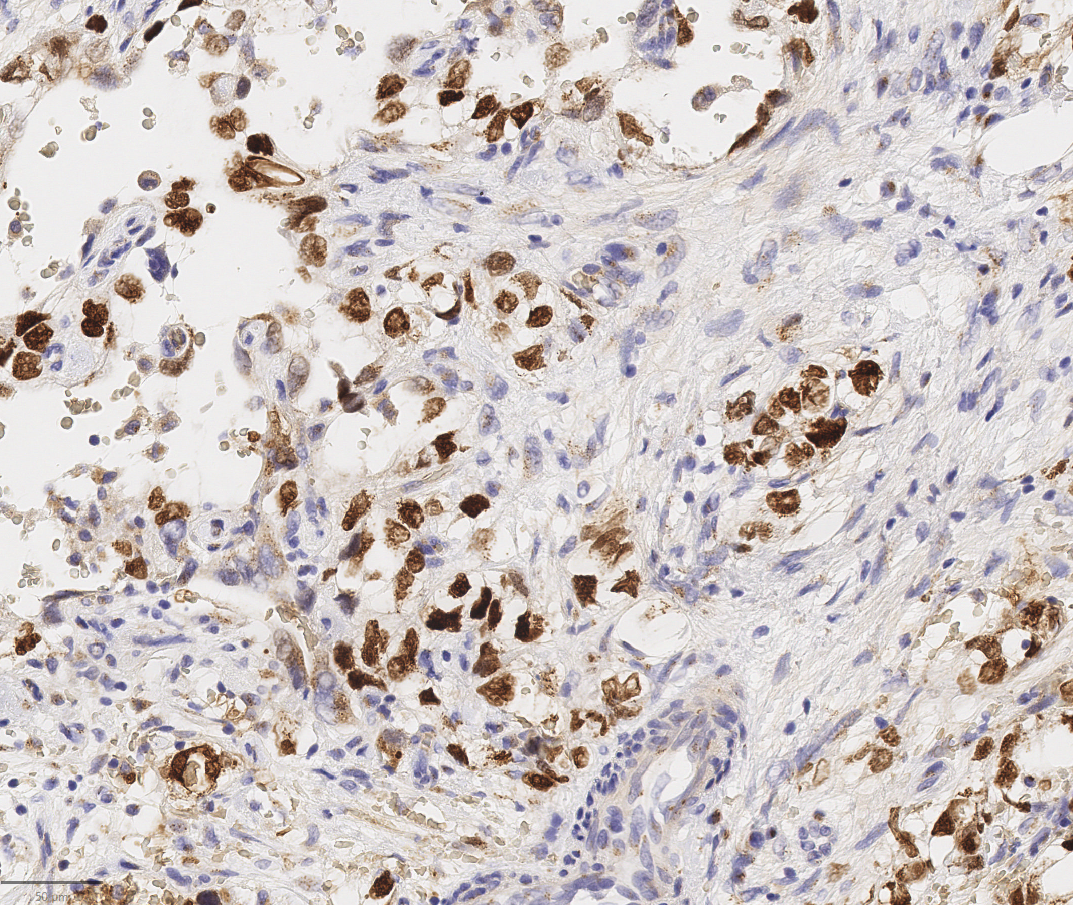
Cd117 c kit stain of mixed malignant germ cell tumor stainin
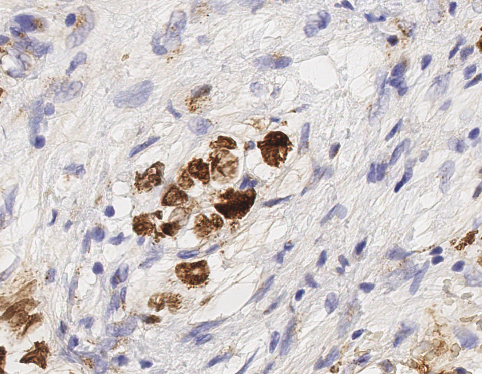
Cd117 c kit stain of mixed malignant germ cell tumor stainin
Identifier cu31924104226240 find matches title general pathoInternet Archive Book Images

Ki67 calculation by qupath in a pure seminoma the colors repCC BY 4.0

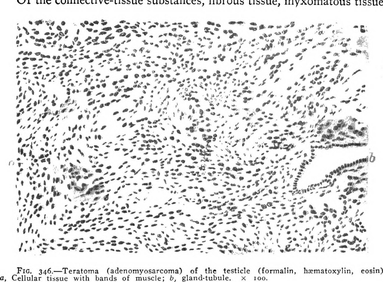
This is the same case presented in image mature cystic teratPublic domain

This 21 year old male was aware of this 10 cm testicular masCC BY 2.0

Mixed germ cell tumor of testis w ruler pathological and hisCC BY 2.0

High magnification micrograph of a mixed germ cell tumour alCC BY-SA 3.0

Intermediate magnification micrograph of a mixed germ cell tCC BY-SA 3.0

Very high magnification micrograph of a mixed germ cell tumoCC BY-SA 3.0

Mixed germ cell tumor containing embryonal carcinoma seminomCC BY-SA 4.0

Most common germ cell tumor of testesCC BY-SA 4.0

Most common germ cell tumor of testesCC BY-SA 4.0

Ccdc188 rna seq data in testicular germ cell tumors versus hCC BY-SA 4.0

Identifier pathologysurgica1895senn find matches title the pNo restrictions
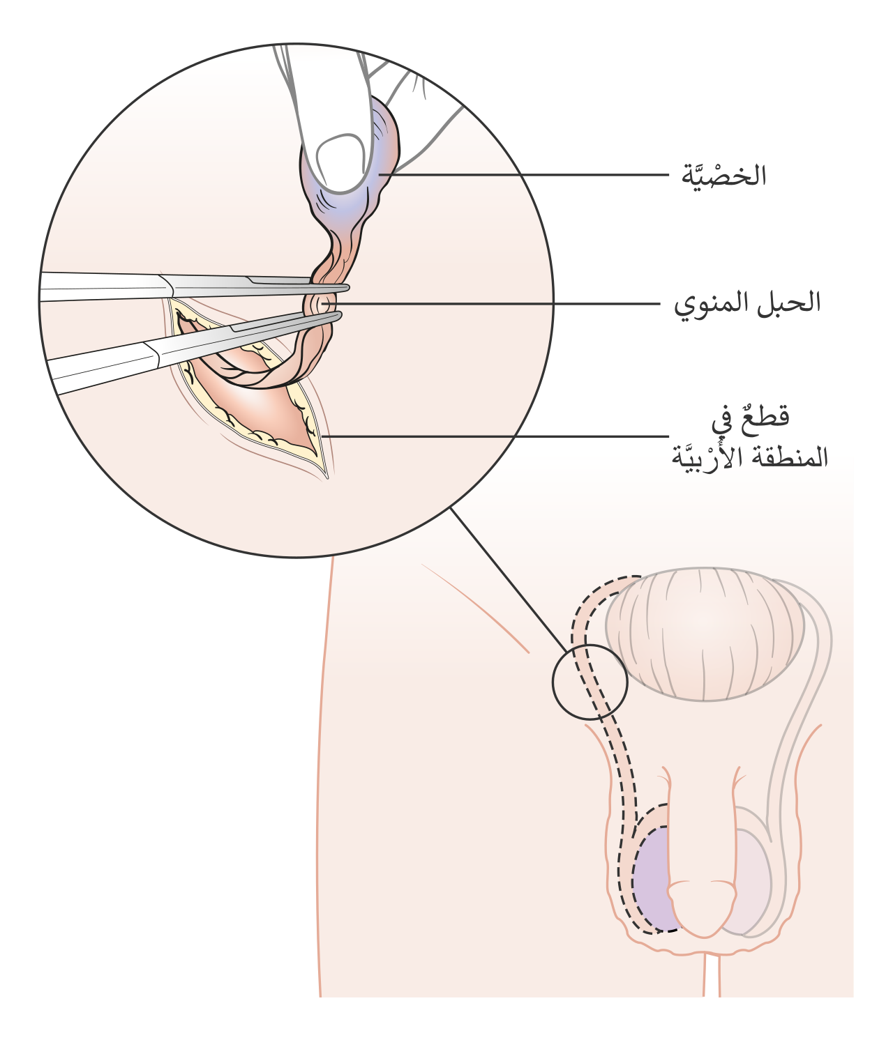
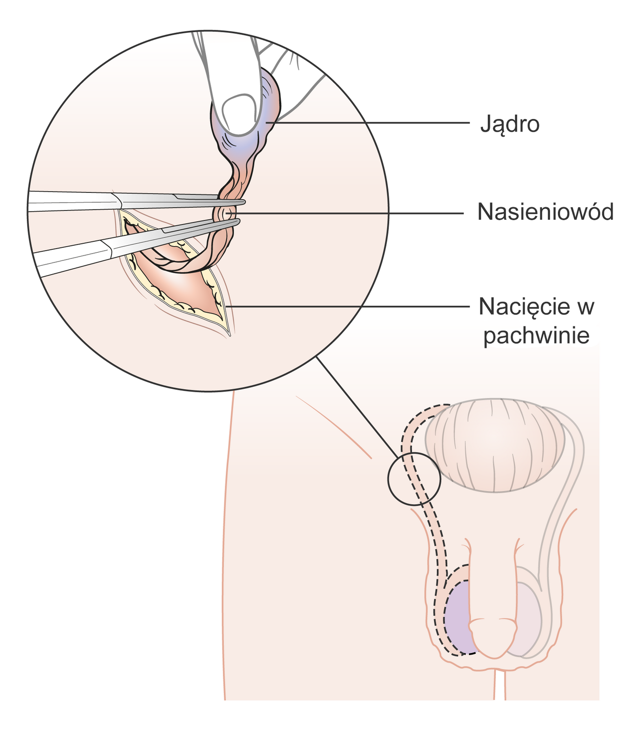
Diagram showing how the testicle is removed orchidectomy

Diagram showing how the testicle is removed orchidectomyCC BY-SA 4.0
Schemat przedstawiaj cy orchidektomi z dost pu pachwinowego
HBP et troubles mictionnels
51 images
Kurdish di temen kaltiy de bi taybet pi t temen 60 saliy proPublic domain

Identifier diseasesofbladde00jone find matches title diseaseNo restrictions

Identifier practiceofsurger00mumf find matches title the praNo restrictions

Balk de luts mei flapbr geCC BY-SA 4.0

De luts in balk in gemeente gaasterl n sleat provincie friesCC BY-SA 3.0

Bomen en straat aan de lutsCC BY-SA 3.0

De lutsCC BY-SA 3.0

De luts bij de kippenburgCC0

De luts netherlandsPublic domain

Depiction of a lady who has a urinary tract infection uti thCC BY-SA 4.0

Panoramic view during sunset of dasht e loot in english emptCC BY-SA 4.0

Hdr panoramic view of approx 180 degrees during sunset of daCC BY-SA 4.0

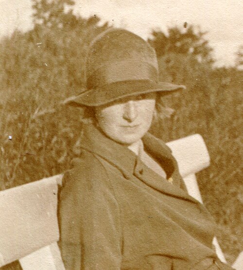
Karin luts arumaa avfotograferad portr ttbild fr n allhems sPublic domain

Karin luts vestlus p rastl unal 1957 1978 tartmusCC BY 4.0
Karin luts vitraa ikavand i 1947 tartmusCC BY 4.0

Karin luts vitraa ikavand iii 1947 tartmusCC BY 4.0
Karin luts vitraa ikavand iv 1947 tartmusCC BY 4.0
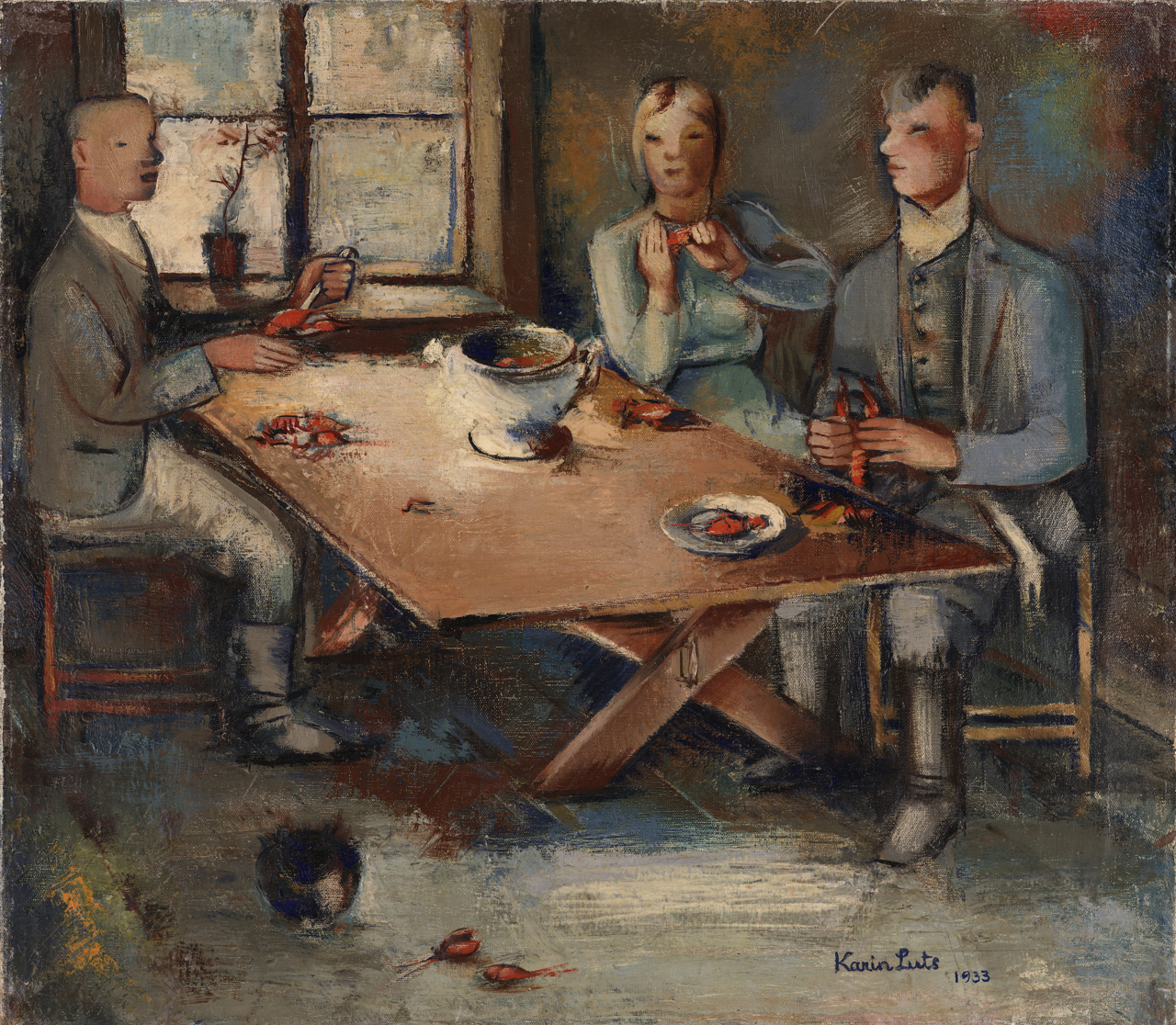
Karin luts v hjas ming 1933 tartmusCC BY 4.0

Karin luts self portrait 1943 tartmusCC BY-SA 4.0
Kunstnik karin luts rannas pingil istumas

Loopbrug over de lutsCC BY-SA 3.0

Siim luts eesti soome maav istlusel veritase staadionilCC BY-SA 4.0

Oskar luts 1910Public domain

Oskar luts 1912Public domain

Oskar luts abikaasa ja pojaga vitebskisPublic domain

Lasgo van links naar rechts david vervoort peter luts evi goCC BY-SA 4.0

Image of estonian filmmaker theodor luts 1896 1980Public domain

Winkels aan de luts in balkCC BY-SA 3.0

A uroflowmeter is a measuring device for measuring the urineCC BY-SA 4.0

Normal uroflowmetryCC BY-SA 3.0

Obstructive uroflowmetryCC BY-SA 3.0

Uroflowmetry uroflow chart and the main quantitative indicatCC BY-SA 4.0

A urologist performing a bipolar turp transurethral resectioCC BY-SA 4.0

This is an example of a causal diagram also known as a web oCC BY-SA 4.0

Title digital rectal exam spanish description examen rectalPublic domain

Kurdish prostat li bin m zdank li p iya tortorik de cih digiPublic domain
Acid phosphatase homotetramer human prostate gland

Identifier americantextbook00howe find matches title an amerNo restrictions
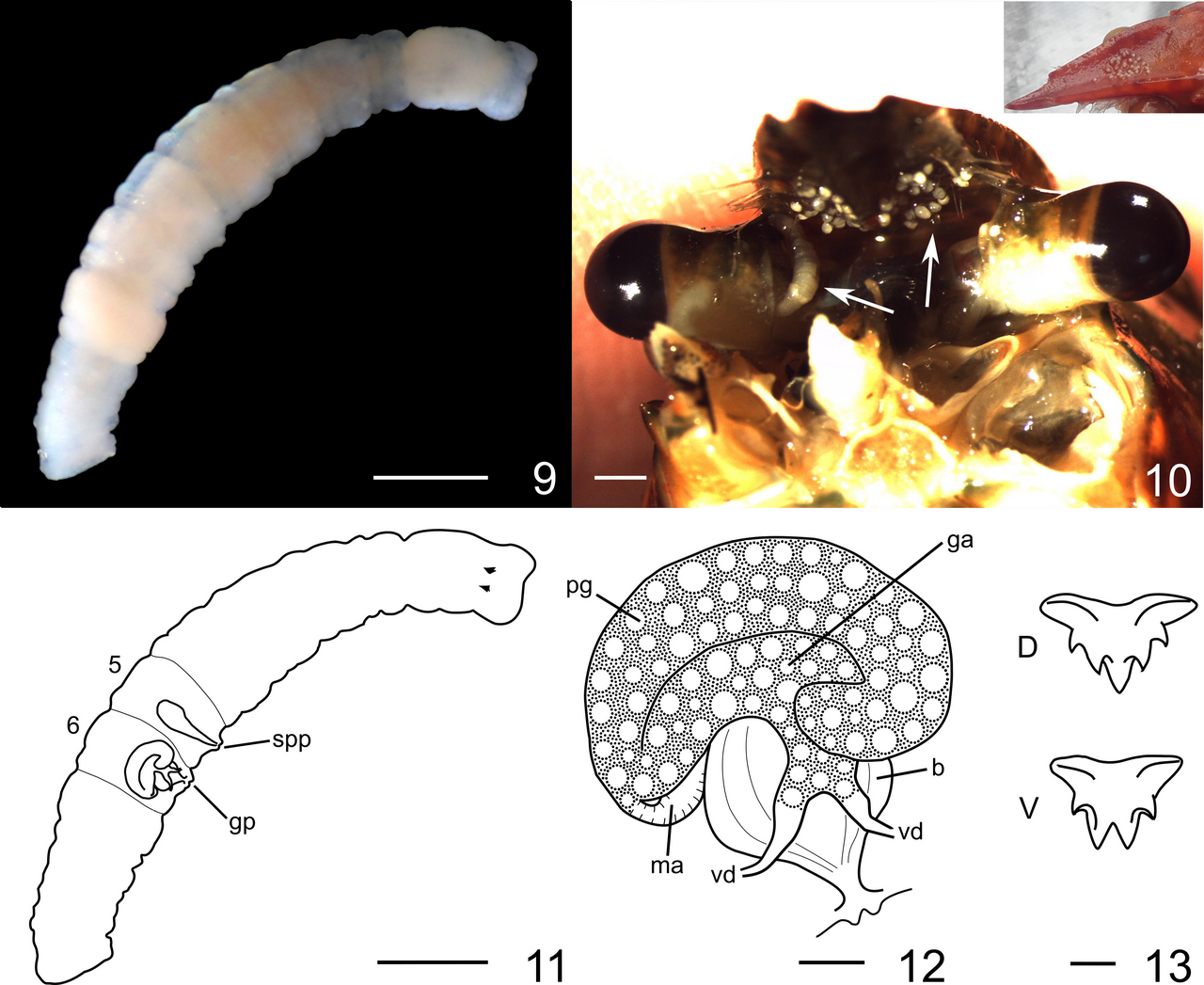
Figures 9 13 cambarincola mesochoreus for abbreviations see Gelder, S.R. (2020). North American Branchiobdellida (Annelida: Clitellata) or Crayfish Worms in France: the most diverse distribution of these exotic ectosymbionts in Europe. Zoosymposia, 17: 121–140.

Figure 3 enchiridium daidai sp nov ichum 5993 holotype schemCC BY 4.0

Superficial surgical anatomy of perineum identifier operativNo restrictions

Figure 2 paraplehnia seisuiae sp nov ichum 5345 holotype sagCC BY 4.0

ProstatePublic domain
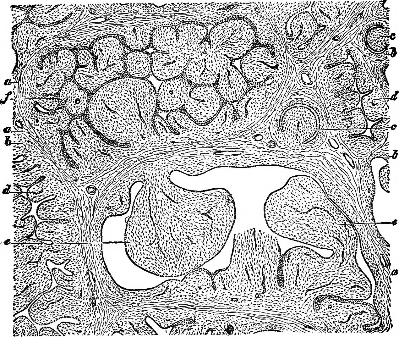
Zones of the prostate
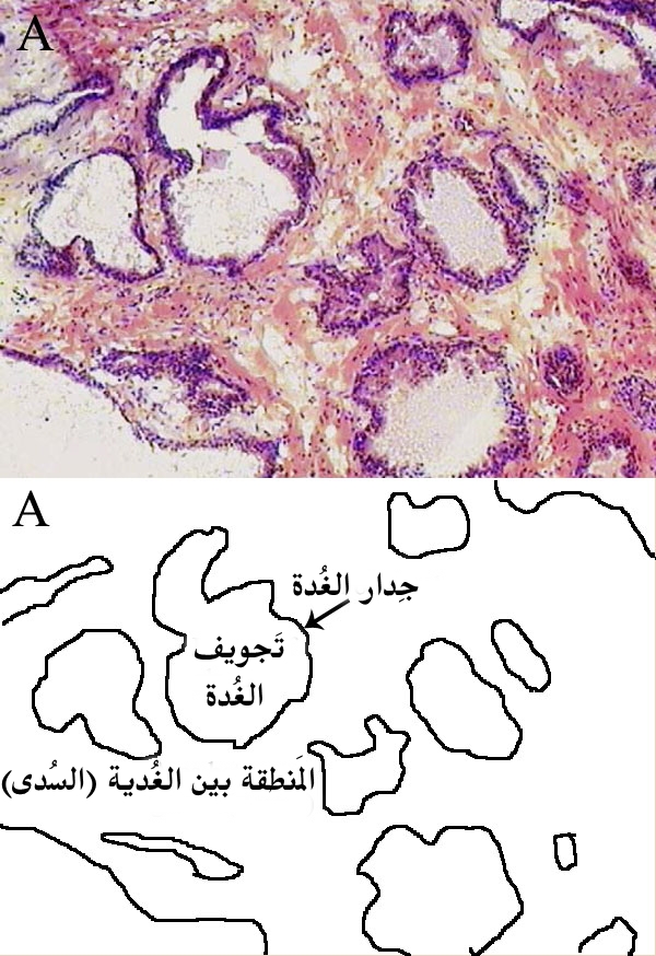
Histological section of prostate gland

Dissection of prostate showing urethra and seminal colliculuCC BY 3.0

Identifier radiographyxrayt00knoxuoft find matches title radNo restrictions

Figure 2 temnocephala ivandarioi sp nov a adult specimen diaCC BY 4.0

Identifier 04801644 5817 emory edu title the pathology and tNo restrictions

Title the cat an introduction to the study of backboned animNo restrictions

Title the chordates identifier chordates00rand find matchesNo restrictions
Infections urinaires
59 images
Bilateral pyelonephritis on plain ct scan as evidenced by faCC BY-SA 4.0

Bacillus calmette guerin bcg induced cystitis showing chroniCC BY-SA 4.0

Bacillus calmette guerin bcg induced cystitis showing chroniCC BY-SA 4.0

Bullous cystitis with prominent edematous broad based papillCC BY-SA 4.0

Bullous cystitis with prominent edematous broad based papillCC BY-SA 4.0

Cystitis cystica showing nests of urothelium with dilated luCC BY-SA 4.0

Cystitis cystica showing nests of urothelium with dilated luCC BY-SA 4.0

Cystitis cystica showing nests of urothelium with dilated luCC BY-SA 4.0

Cystitis glandularis intestinal type showing mucin secretingCC0

Cystitis glandularis intestinal type showing mucin secretingCC0

Eosinophilic cystitis showing edematous and chronically inflCC BY-SA 4.0

Eosinophilic cystitis showing prominent eosinophils in the mCC BY-SA 4.0

Eosinophilic cystitis showing edematous and chronically inflCC BY-SA 4.0

Eosinophilic cystitis showing edematous and chronically inflCC BY-SA 4.0

Follicular cystitis showing numerous lymphoid follicles in tCC BY-SA 4.0

Follicular cystitis showing numerous lymphoid follicles in tCC BY-SA 4.0

Follicular cystitis showing numerous lymphoid follicles in tCC BY-SA 4.0
Histopathology of radiation cystitis with atypical stromal c

Papillary cystitis with finger like projections and ectaticCC BY-SA 4.0

Papillary cystitis with finger like projections and ectaticCC BY-SA 4.0

Papillary cystitis with finger like projections and ectaticCC BY-SA 4.0

Polypoid cystitis showing broad polypoid growth prominent blCC BY-SA 4.0

Polypoid cystitis showing broad polypoid growth prominent blCC BY-SA 4.0

Polypoid cystitis showing broad polypoid growth prominent blCC BY-SA 4.0

Surgical anatomy of the perineum identifier textbookofoperatNo restrictions

These bacteria were cultured from a urine sample from a womaCC BY-SA 3.0

Escherichia coli gram negative rod off a culture plate fromCC BY-SA 4.0

Uti agar is a chromogenic medium for identification and diffCC BY-SA 4.0

Identifier annualreport192627univ find matches title annualNo restrictions

Identifier urinaryanalysi00heit find matches title urinary aNo restrictions

Identifier diseasesofwomenc00herm find matches title diseaseNo restrictions
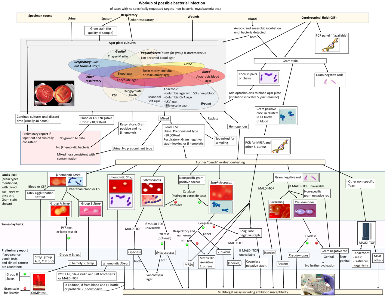
Example of a workup algorithm of possible bacterial infectio

Identifier filariasanguinis00mans find matches title the filNo restrictions

Identifier handbookofmedic00rile find matches title handbookNo restrictions

9 bucuresti romania parcul cismigiu in ultima zi de iarna 20CC BY-SA 4.0

Rectum much distended artificially identifier manualofmodernNo restrictions

Identifier newmanualofsurge00ochs find matches title a new mNo restrictions

Photo of the sacrificial altar dedicated to cautes one of thCC BY 4.0

Identifier americantextbook00bang find matches title an amerNo restrictions

Identificatietitel s arme ouders rijke kinderen rijke kinderCC0

Particolare del momento ai cauti sulla facciata della chiesaCC BY-SA 4.0
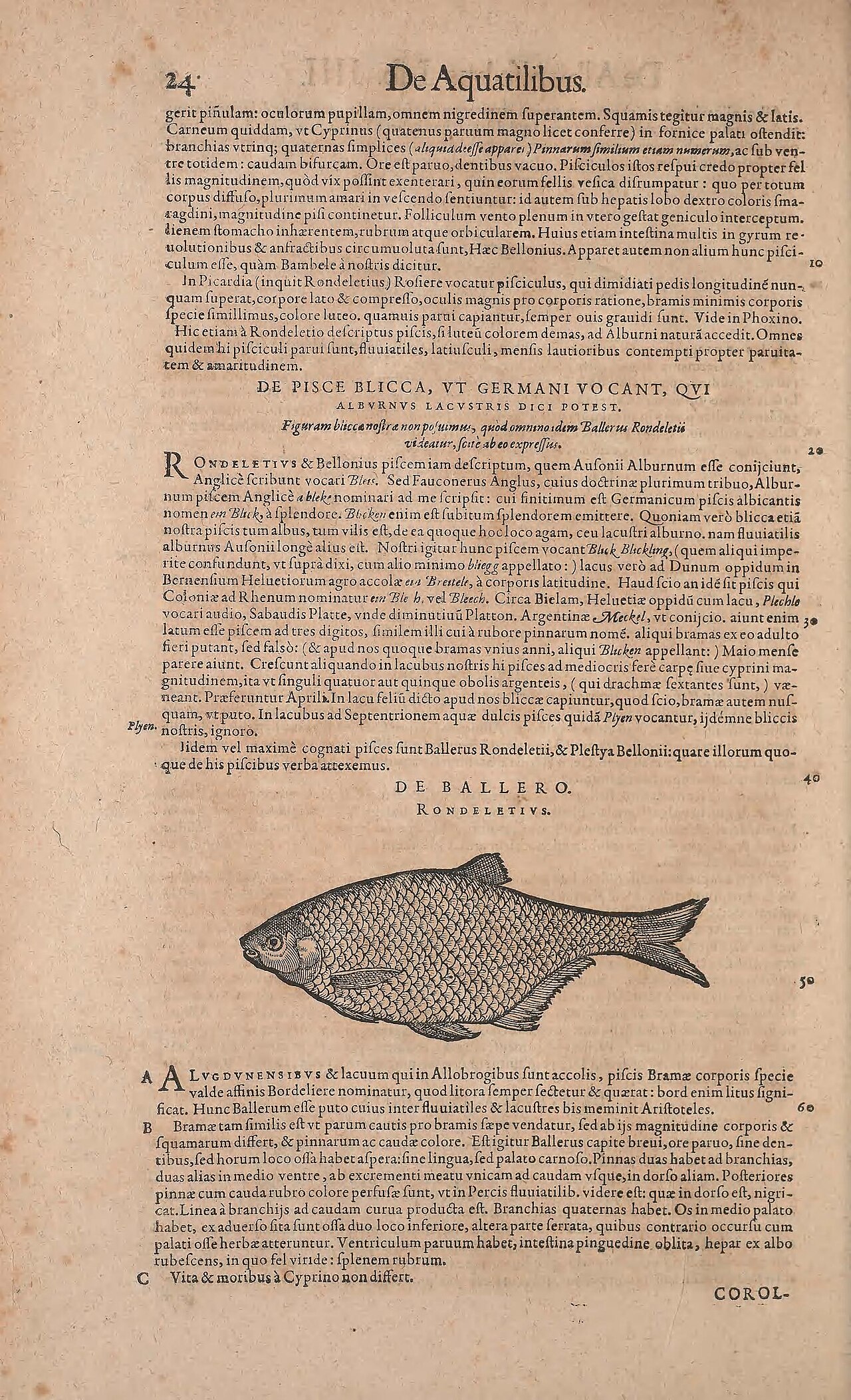
24 deaquatilibus gcrit pinulam oculorum pupillam omncm nigre Cambier, Andreas

Identificatietitel s dronken silenus vinum cautis innocuum tCC0

Identificatietitel s dronken silenus vinum cautis innocuum tCC0
Identifier floridaitshistor00fair find matches title floridaInternet Archive Book Images

Identifier gynaecologyforst00eden find matches title gynaecoNo restrictions

Identifier mansmissiononear00kahn find matches title man s mNo restrictions

Identifier mansmissiononear00kahn find matches title man s mNo restrictions

Identifier monumentumextrem00veran find matches title monumeNo restrictions

Identifier monumentumextrem00veran find matches title monumeNo restrictions

Identifier comprehensivehis01beve title a comprehensive histNo restrictions

Postredirectget doublesubmitsolutionLGPL

Identifier transactionsofwe1917west find matches title transNo restrictions

In a urine microscopy sample from a patient with a fungal utCC BY-SA 4.0

General view of clinical technologist mullin chiong measurinNo restrictions

When you have a urine sample with packed pus cells and bacteCC BY-SA 4.0

Identifier plainhometalkabo00foot find matches title plain hNo restrictions

Identifier examinationofuri00fish find matches title the exaNo restrictions

Identifier floridaitshistor00fair find matches title floridaInternet Archive Book Images
Andrologie
78 images
3d medical animation still showing the inability to developCC BY-SA 4.0

3d medical animation still showing the inability to developCC BY-SA 4.0

Normal erection l vs erectile dysfunction rCC BY-SA 4.0

Left testicular seminoma varicocele ultrasound imageCC BY-SA 3.0

Scrotal ultrasonography of an intratesticular varicoceleCC BY 3.0

Scrotal ultrasonography of varicoceleCC BY 3.0
Varicocele and testis in ultrasound transverse imageCC BY-SA 3.0

Varicocele and testis in ultrasound transverse imageCC BY-SA 3.0

Erect penis of 60 year old male with peyronie s diseaseCC0

Plaques in penis shaft possibly due to injury cause the peniCC BY-SA 4.0

Curved penis due to peyronies diseaseCC BY-SA 4.0

Erected human penis bent to the side 90 degrees curvature peCC BY-SA 4.0

Erected human penis bent to the side 90 degrees curvature peCC BY-SA 4.0

Curved and bent penis due to peyronies diseaseCC BY-SA 4.0

Curved and bent penis due to peyronies diseaseCC BY-SA 4.0

Curved and bent penis due to peyronies diseaseCC BY-SA 4.0

Peyronie s disease in an erect penis little sharp bend to leCC BY-SA 4.0

Peyronie s disease in an erect penis little sharp bend to leCC BY-SA 4.0

Peyronie s disease in an erect penis little sharp bend to leCC BY-SA 4.0

Peyronie s disease in an erect penis little sharp bend to leCC BY-SA 4.0

Peyronie s disease in an erect penis little sharp bend to leCC BY-SA 4.0
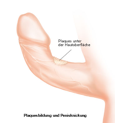
Peyronie s disease nodules so called plaques under the skin Medical Art Service

Erected curved human penis bent crooked peyroniesCC BY-SA 4.0
Erection development of a penis exhibiting signs of peyronie

Abnormal upward curvature of erect penis caused by peyronieCC BY-SA 4.0

Erect penis of 60 year old male with peyronie s diseaseCC0

Chronic peyronie s disease penis from topCC BY-SA 4.0

This ultrasound depicts both normal and abnormal anatomy inCC BY-SA 3.0

This is an example of a patient with congenital from birth pCC BY-SA 3.0

Penile curvature typical of an advanced stage of peyronie sPublic domain

Peyronie s disease curved penis penis curvature penis deformCC BY-SA 4.0

Before left and after middle onset of peyronie s disease cauCC0

Peyronie s diseasePublic domain

Fig 1 schematic diagram showing classification of hydroceleCC BY 4.0

Wan shijia chuanguang si jiyao digest of knowledge handed doCC BY 4.0

Wan shijia chuanguang si jiyao digest of knowledge handed doCC BY 4.0

Wan shijia chuanguang si jiyao digest of knowledge handed doCC BY 4.0

Wan shijia chuanguang si jiyao digest of knowledge handed doCC BY 4.0

Wan shijia chuanguang si jiyao digest of knowledge handed doCC BY 4.0

Wan shijia chuanguang si jiyao digest of knowledge handed doCC BY 4.0

Wan shijia chuanguang si jiyao digest of knowledge handed doCC BY 4.0

Wan shijia chuanguang si jiyao digest of knowledge handed doCC BY 4.0

Wan shijia chuanguang si jiyao digest of knowledge handed doCC BY 4.0

Wan shijia chuanguang si jiyao digest of knowledge handed doCC BY 4.0

Wan shijia chuanguang si jiyao digest of knowledge handed doCC BY 4.0

Wan shijia chuanguang si jiyao digest of knowledge handed doCC BY 4.0

Wan shijia chuanguang si jiyao digest of knowledge handed doCC BY 4.0

Wan shijia chuanguang si jiyao digest of knowledge handed doCC BY 4.0

Wan shijia chuanguang si jiyao digest of knowledge handed doCC BY 4.0

Wan shijia chuanguang si jiyao digest of knowledge handed doCC BY 4.0

Wan shijia chuanguang si jiyao digest of knowledge handed doCC BY 4.0

Wan shijia chuanguang si jiyao digest of knowledge handed doCC BY 4.0

Wan shijia chuanguang si jiyao digest of knowledge handed doCC BY 4.0

Wan shijia chuanguang si jiyao digest of knowledge handed doCC BY 4.0

Wan shijia chuanguang si jiyao digest of knowledge handed doCC BY 4.0

Wan shijia chuanguang si jiyao digest of knowledge handed doCC BY 4.0

Fig 9 a diagram showing the pattern of spermatogenesis in gCC BY 4.0

Fig 3 a schematic diagram showing a germinal chamber of theCC BY 4.0

Identifier textbookofdent00noye find matches title a text boNo restrictions

Proceso de espermatog nesisCC BY-SA 3.0

Identifier journalofexperim18broo find matches title journalNo restrictions
Spermatogenesis

Spermatogenesis
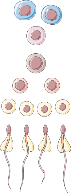
Spermatogenesis
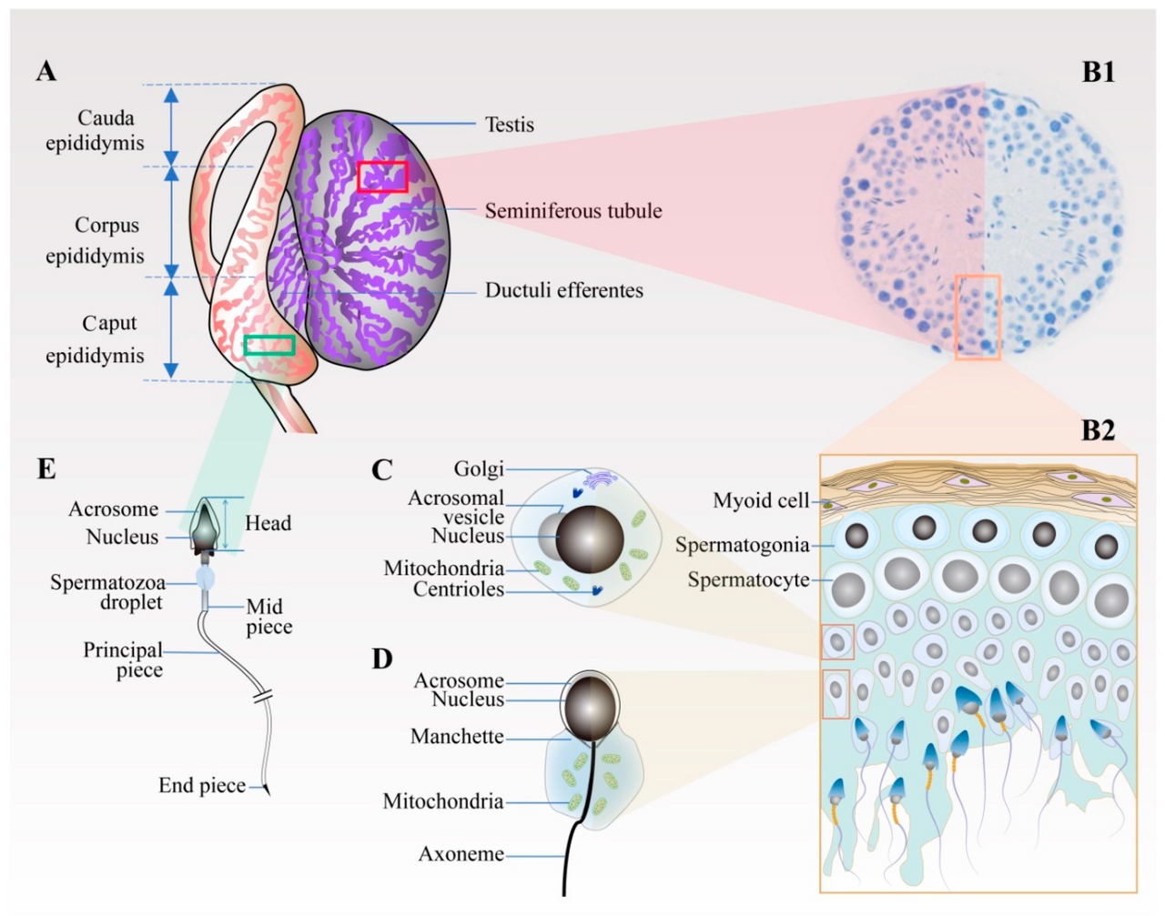
Figure 1 schematic diagram of spermiogenesis in the testis a Duan, C.

The cycle of the seminiferous epithelium shown within a crosCC BY-SA 4.0

Scheme of the spermatogenesisCC BY-SA 4.0

Schema fasi spermatogenesiCC0

Figure 3 left a diagram depicting the cell cycle progressionCC BY 4.0

Diagram of the creation over sperm via the process spermatogCC BY-SA 4.0

Diagram of spermatogenesis labelled in gallician languageCC0

Kurdish ji xaney n d plo d bi dabe b na miyoz diristkirina sCC BY-SA 4.0

Scheme of spermatogenesis with czech descriptionCC BY 3.0 cz

Simple grayscale diagram with not labels comparing mitosis oCC BY-SA 3.0
Title the physiology of the domestic animals a text book forInternet Archive Book Images

The process of spermatogenesisPublic domain

Kurdish ji destp ka temen p gih t n heta dawiya jiyan di laCC BY 4.0

The calcium homeostasis of modern sperm cells looks very simCC BY 4.0
Incontinence et urodynamique
38 images
3d medical animation still showing normal urinary bladder lCC BY-SA 4.0

A diagram of the catheters used in cytometryPublic domain

A diagram of the pelvic floor musclesCC BY 4.0

Adult swim diaper for fecal incontinenceCC0

Burch suspension for urinary incontinencePublic domain

The result of fecal incontinencePublic domain

ImageCC BY-SA 4.0

Incontinence pad for men holding penis within the slitCC BY-SA 3.0

Incontinence pad for men holding penis sticking on briefsCC BY-SA 3.0

Incontinence pad for men packageCC BY-SA 3.0

Incontinence pad for men packageCC BY-SA 3.0

Incontinence pad for menCC BY-SA 3.0

Incontinence pad for men without penis holding sticking on bCC BY-SA 3.0

Incontinence pad for womenCC BY-SA 3.0

Incontinence pad for women sticking on pantiesCC BY-SA 3.0

Incontinence pad for women packageCC BY-SA 3.0

Incontinence pad for women packageCC BY-SA 3.0

This sanitary bin was in a women s toilet in the uk howeverCC BY-SA 4.0
3d medical animation still showing normal urinary bladder lCC BY-SA 4.0

Burch procedureCC BY-SA 4.0

Version 8 25 from the textbook openstax anatomy and physioloCC BY 4.0

Identifier manualofgyncol00skeeuoft find matches title a manNo restrictions

Identifier manualofgyncol00skeeuoft find matches title a manNo restrictions

Healthcare professional educating on pelvic structuresCC BY 4.0

Male pelvic floorCC BY-SA 4.0

The pelvic floor is a group of muscles and tissues that suppPublic domain
A diagram of the pelvic floor musclesCC BY 4.0

Physiotherapist explaining pelvic floor functionCC BY 4.0

Identifier diagnosistreatmecros find matches title the diagnNo restrictions

This is not a good picture as there is no emg tracing strictPublic domain
A diagram of the catheters used in cytometryPublic domain

Bladder is innervated peripherally and centrally lesion of wCC BY 4.0

Comparison of extended release formulations of oxybutynin anCC0

Skeletal formula of solifenacin a muscarinic antagonist usedPublic domain

Skeletal formula of trospium chloride a muscarinic antagonisPublic domain

Identifier treatiseonobstet00davi find matches title a treatNo restrictions

Identifier diseasesofchildr00grah find matches title diseaseNo restrictions
Identifier postoperativetr00mors find matches title postoperInternet Archive Book Images
Urologie pediatrique
14 images
A color coded diagram drawn over a photo showing vulvar hypoCC0

ImageCC BY 4.0
Identifier generaldentalpat00ende find matches title generalInternet Archive Book Images
Identifier plasticsurgeryit00davi find matches title plasticInternet Archive Book Images

A diagram of hypospadias and epispadias as may be seen in aCC0

Grades 1 vesico ureteric reflux shown on right of the diagraCC BY-SA 4.0

Grades 1 to 5 vesico ureteric reflux shown on right of eachCC BY-SA 4.0

Grades 2 vesico ureteric reflux shown on right of the diagraCC BY-SA 4.0

Grades 3 vesico ureteric reflux shown on right of the diagraCC BY-SA 4.0

Grades 4 vesico ureteric reflux shown on right of the diagraCC BY-SA 4.0

Grades 5 vesico ureteric reflux shown on right of the diagraCC BY-SA 4.0

Bilateral dilatation of the ureters due to vesicoureteric reCC BY 4.0

Identifier generaldentalpat00ende find matches title generalInternet Archive Book Images

Identifier generaldentalpat00ende find matches title generalInternet Archive Book Images
Transplantation renale
46 images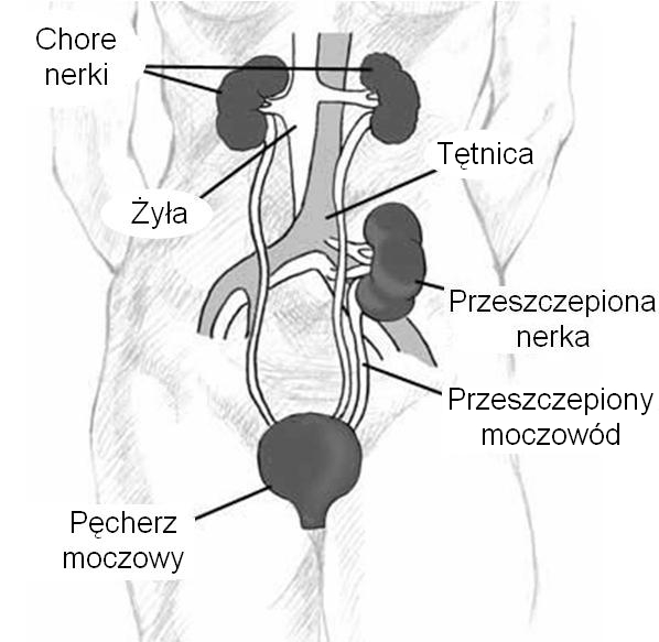
Kidney location after transplantation
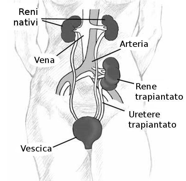
Kidney location after transplantation adapted from the origi

Trasplantament renalCC BY-SA 3.0

Siirtomunuaisen sijainti munuaisensiirrossaCC BY 3.0

Kidney location after transplantation adapted from the origiPublic domain

Kidney location after transplantationCC BY 3.0

Kidney location after transplantation inscriptions in bulgarPublic domain

Diseases and conditions where stem cell treatment is promisiPublic domain
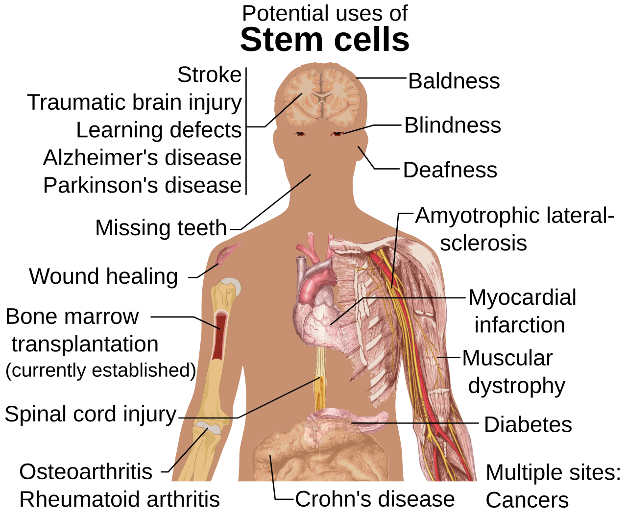
Diseases and conditions where stem cell treatment is promisi

Descriptive graph of kidney transplantCC BY-SA 4.0

The patient is a 39 y o female with a h o lupus who is postCC BY-SA 4.0
Image

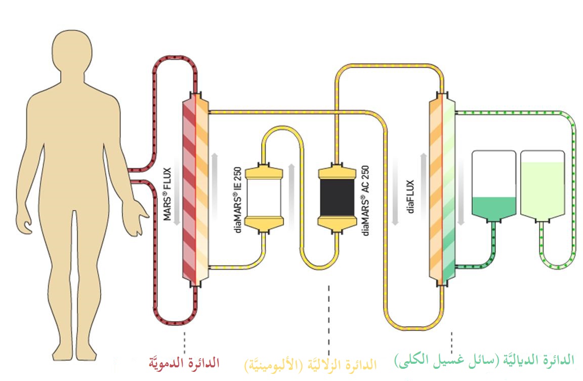
Albumin dialysis circuitCC BY-SA 3.0

ImageCC BY-SA 3.0

Dialysis diagramPublic domain
This is an illustration of the life cycle of the parasitic a

Diagram of osmosis in a u shaped tube through a dialysis memCC0
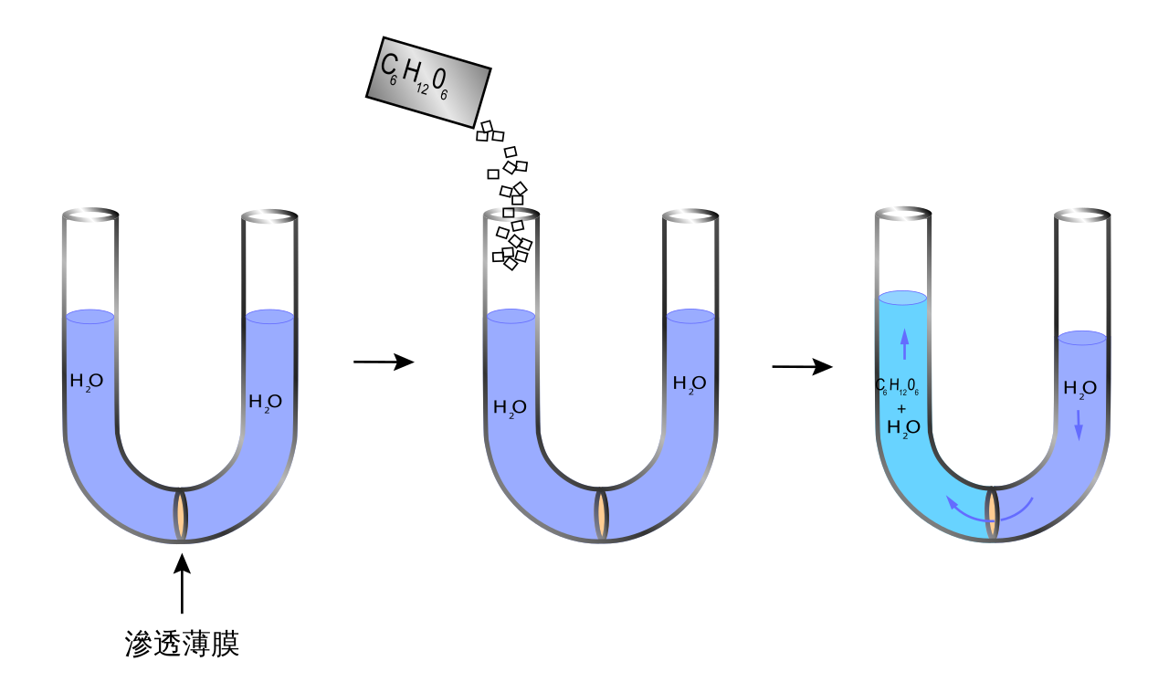
Diagram of osmosis in a u shaped tube through a dialysis mem

Diagram of osmosis before and after through a semi permeableCC0

If you can improve this diagram please do propose things onCC BY 4.0

A dialysis machine is used for implementing and monitoring hCC BY-SA 4.0

Exhibit in the tekniska museet stockholm sweden this work isCC0

Hcch s hdr head rn operating a new hemodialysis machinePublic domain

Nih instituto nacional de la diabetes y las enfermedades digCC BY-SA 4.0

Simplified hemodialysis circuitCC BY-SA 4.0

Esquema de la hemodi lisiCC BY-SA 3.0
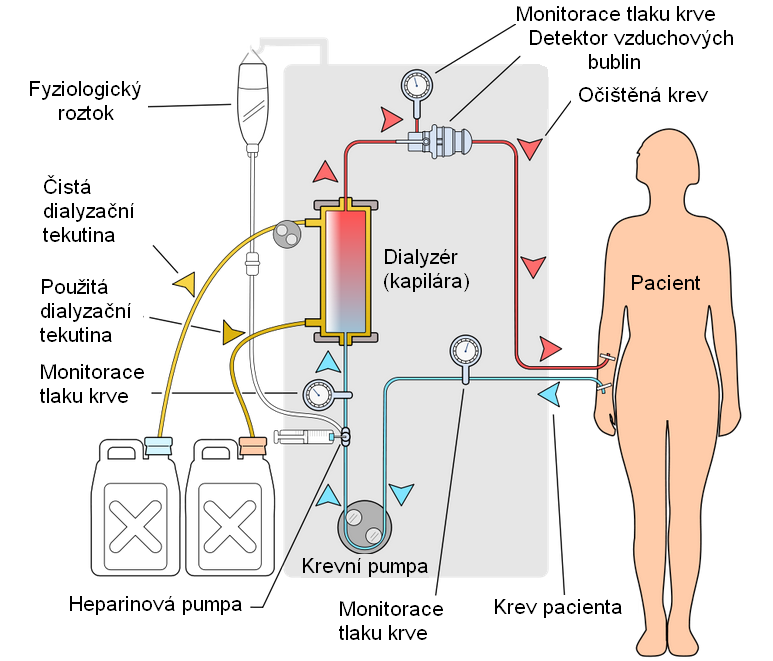
Simplified hemodialysis circuit

Simplified hemodialysis circuitCC BY-SA 3.0

Simplified hemodialysis circuitCC BY 3.0

Simplified hemodialysis circuitCC BY-SA 3.0

Simplified hemodialysis circuitCC BY-SA 3.0

Simplified hemodialysis circuitCC BY 3.0

Hemodial zis v zlataCC BY-SA 4.0

Hemodial zis v zlatos br zol saCC BY-SA 4.0

Blood is taken into the machine also known as hemodialysis eCC BY-SA 3.0

Hemodialysis graft used in hemodialysysPublic domain

InnovaCC BY-SA 3.0

Schematic of a hemodialysis circuitPublic domain

A hemodialysis machine in linkou chang gung memorial hospitaCC BY-SA 4.0

A patient is lying in a hospital bed in an intensive care unCC BY-SA 4.0

Patient receiving dialysisCC0

Arm of patient receiving dialysisCC0

U s air force col robert noll 349th medical squadron commandPublic domain

U s air force col robert noll 349th medical squadron commandPublic domain

Old men is having an diagnostic testCC BY-SA 3.0

Patient receiving dialysisPublic domain
Techniques chirurgicales et endoscopie
47 images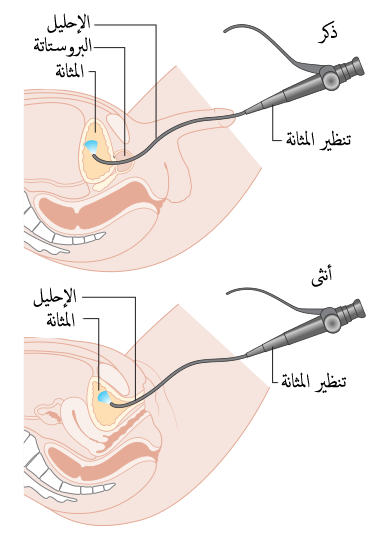
Diagram showing a cystoscopy for a man and a woman

Diagrama d una cistosc pia en un home i en una donaCC BY-SA 3.0

Diagram showing a cystoscopy for a man and a womanCC BY-SA 4.0
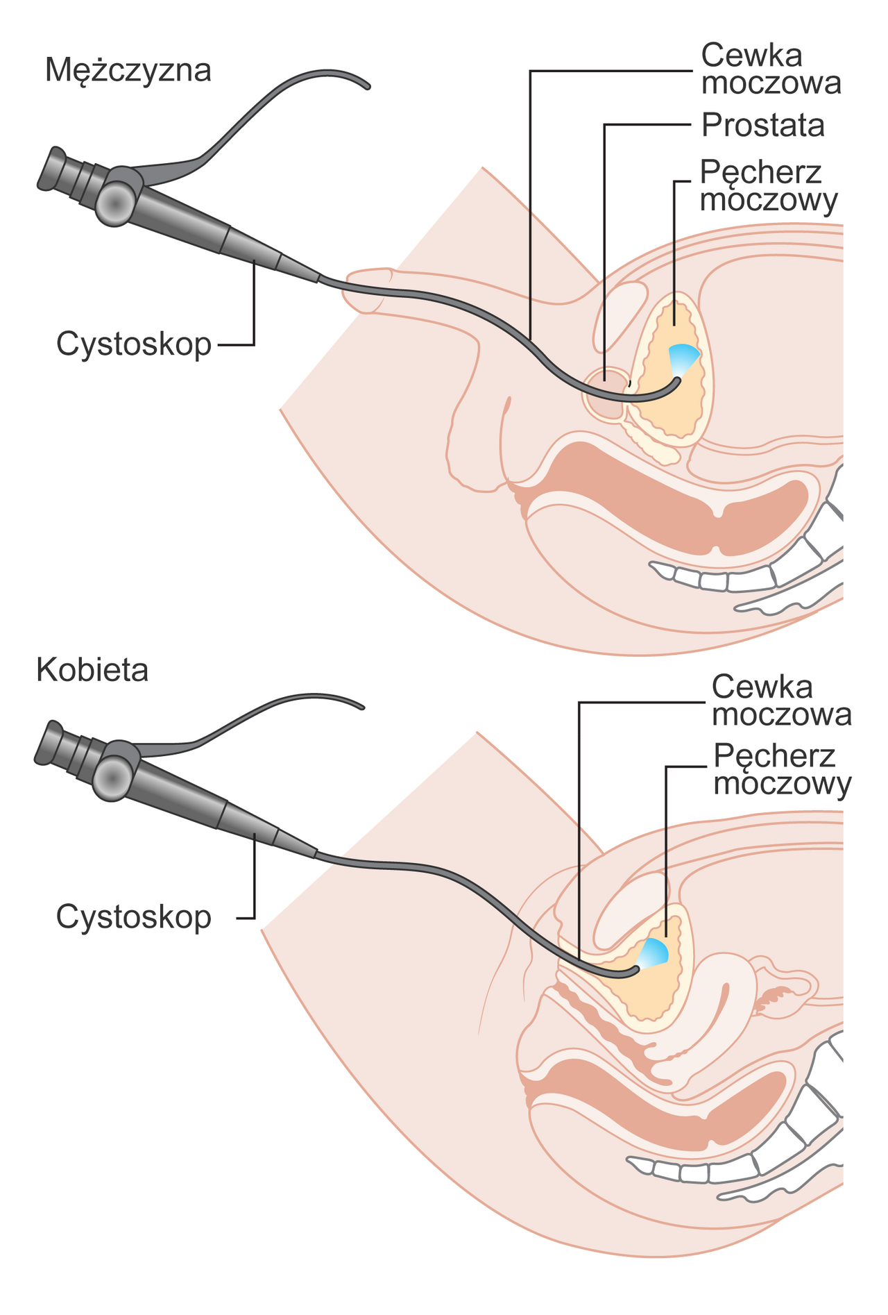
Diagram przedstawiaj cy cystoskopi u m czyzny i kobiety

ImageCC BY-SA 4.0

Diagram showing a cystoscopy for a man and a woman in bulgarCC BY 4.0

Diagram showing laparoscopic surgery for kidney cancerCC BY-SA 4.0

People watching 3d projection of chirurgical operation by acCC BY 3.0

Lt col dr thomas shaak 81st medical support squadron clinicaPublic domain

Lt col dr thomas shaak 81st medical support squadron clinicaPublic domain

Medical building on i 10 service road across from lakeside hCC BY-SA 4.0

Medical building on i 10 service road across from lakeside hCC BY-SA 4.0

Col debra lovette 81st training wing commander receives a brPublic domain

Second lt nina hoskins 81st surgical operations squadron opePublic domain

Col debra lovette 81st training wing commander operates a roPublic domain

A surgeon at the columbia hernia center operates on a patienCC BY-SA 4.0

Dr yugal mishra heart surgeon in india present appointment dCC BY-SA 3.0

External aspect of the operative field davinci robotic systeCC BY 4.0

A laparoscopic robotic surgery machine patient side cart ofCC BY-SA 3.0

Iss070e097708 feb 21 2024 nasa astronaut and expedition 70 fPublic domain

Robotic surgery ad at manipal hospital lift door each robotCC BY-SA 3.0

Dr sudhir kumar rawal during a robotic surgeryCC0

William beaumont army medical center performed the first robPublic domain

William beaumont army medical center performed the first robPublic domain

Medical personnel place sterilized covers on the arms of thePublic domain

Small articulating instruments demonstrate the precision andPublic domain

Medical personnel at william beaumont army medical center pePublic domain

Dr samuel cancel rivera an ob gyn physician looks through aPublic domain

A surgeon s view through a 3d imaging system displays articuPublic domain

Medical personnel at william beaumont army medical center pePublic domain

William beaumont army medical center performs the first roboPublic domain

Plotting of temperature pulse and respiration identifier treNo restrictions
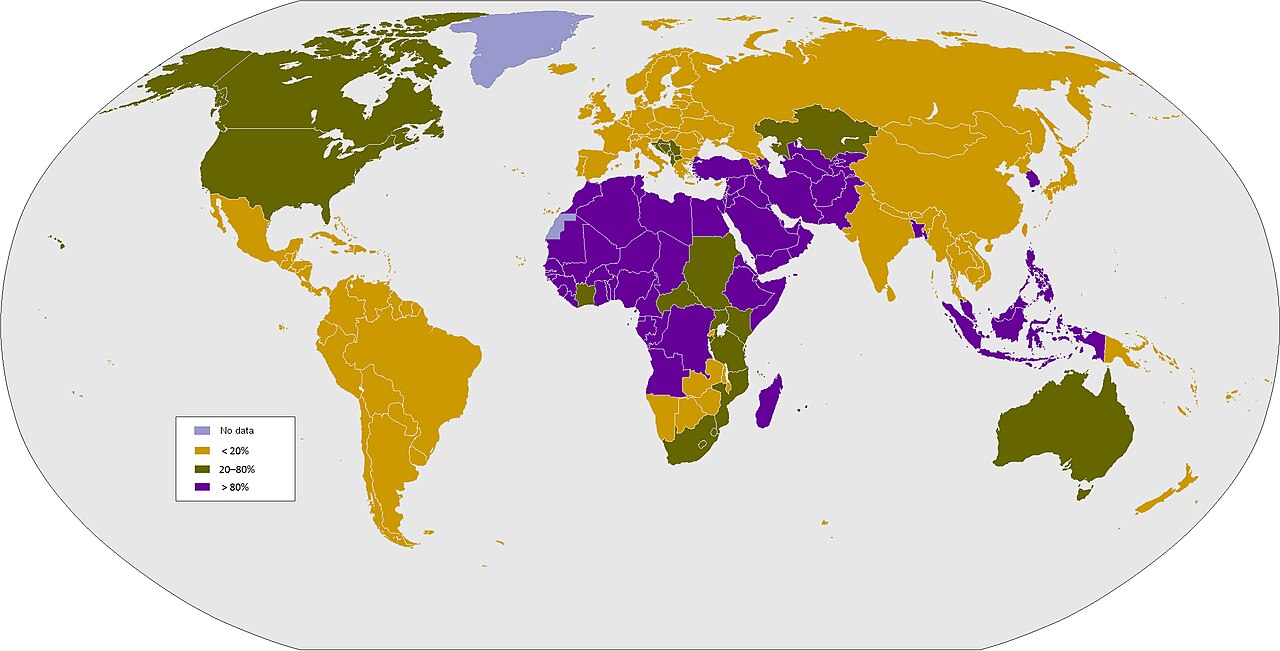
Map showing percentage of males circumcised by country
Image

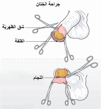
Line drawing diagram of circumcision with hemostats and scisCC BY-SA 3.0

200813 n da693 1148 san diego aug 13 2020 capt sean stroup aPublic domain

Instruments be used by laparoscopic hernia operationCC BY-SA 3.0

The wings hospital s operating theater wings hospital ahmedaCC BY-SA 3.0

Hm3 najii thomas assists lt cmdr matthew bradley chief of trPublic domain

U s air force capt kate hanel 88th surgical operation squadrPublic domain

U s air force major richard knight center diplomate of the aPublic domain

Diagram showing a urinary catheter in a manCC BY-SA 4.0

Diagram showing a urinary catheter in a man who has had hisCC BY-SA 4.0

Diagram showing a urinary catheter in a womanCC BY-SA 4.0

Diagram showing self catheterisation of a urinary diversionCC BY-SA 4.0

Side view diagram of male urinary tract with foley catheterPublic domain
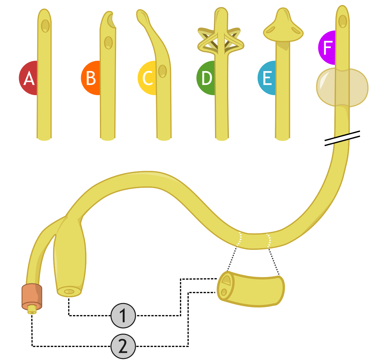
The type of urinary catheters legend a simple urethral cathe